. 2006 Nov;128(5):569-81.
doi: 10.1085/jgp.200609638. Epub 2006 Oct 16.

Detection of the opening of the bundle crossing in KcsA with fluorescence lifetime spectroscopy reveals the existence of two gates for ion conduction

Affiliations

Detection of the opening of the bundle crossing in KcsA with fluorescence lifetime spectroscopy reveals the existence of two gates for ion conduction

Rikard Blunck et al. J Gen Physiol. 2006 Nov.

Abstract

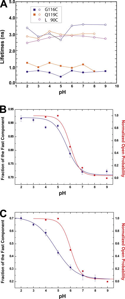
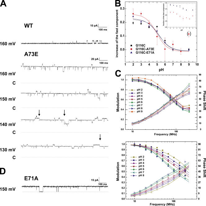
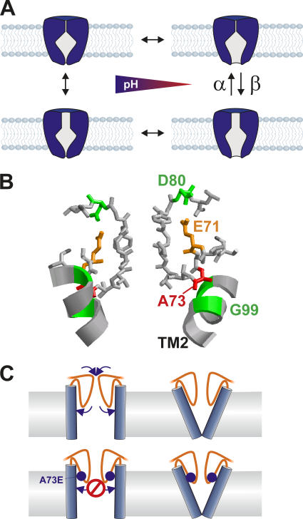
The closed KcsA channel structure revealed a crossing of the cytosolic ends of the transmembrane helices blocking the permeation pathway. It is generally agreed that during channel opening this helical bundle crossing has to widen in order to enable access to the inner cavity. Here, we address the question of whether the opening of the inner gate is sufficient for ion conduction, or if a second gate, located elsewhere, may interrupt the ion flow. We used fluorescence lifetime measurements on KcsA channels labeled with tetramethylrhodamine at residues in the C-terminal end of TM2 to report on the opening of the lower pore region. We found two populations of channels with different fluorescence lifetimes, whose relative distribution agrees with the open probability of the channel. The absolute fraction of channels found with an open bundle crossing is too high to explain the low open probability of the KcsA-WT channel. We found the same distribution as in the WT channel between open and closed bundle crossing for two KcsA mutants, A73E and E71A, which significantly increase open probability at low pH. These two results strongly suggest that a second gate in the ion permeation pathway exists. The location of the mutations A73E and E71A suggests that the second gate may be the selectivity filter, which resides in an inactivated state under steady-state conditions. Since the long closed times observed in KcsA-WT are not present in KcsA-A73E or -E71A, we propose that KcsA-WT remains predominantly in a state with an open bundle crossing but closed (inactivated) second gate, while the mutations A73E and E71A sharply decrease the tendency to enter in the inactivated state, and as a consequence, the second gate is predominantly open at steady state. The ability to monitor the opening of the bundle crossing optically enables the direct recording of the movement of the pore helices while the channel is functioning.


Figures

Figure 1.
Figure 1.
Principle of lifetime measurements in the frequency domain. (A) If a fluorophore absorbs a photon (hν) it is excited to the S1 state. Each fluorophore has a characteristic dwell-time τ in the S1 state before it relaxes back to the ground state S0. This can occur by emitting a photon (red) by nonradiative deexcitation (knr) or by quenching (q). (B) If the amplitude of the excitation light (blue) is sinusoidally modulated, the emission will follow this time course with a time delay (due to the dwell time in S1), resulting in the phase shift ΔΦ. Due to a low-pass filtering effect, the amplitude of the emission is reduced (ΔM). (C) Labeling efficiency: comparison of labeling of single-cysteine mutant and WT channel. The labeling ratio of mutant to WT is ∼13.
Figure 2.
Figure 2.
(A and B) Lifetime measurements of TMR-maleimide (0.2 μM) in different solvents (A) and at different pH in a 0.5 mg/ml lipid vesicles solution (B). Shown are the data (symbols) as well as the fits (lines). The fit results of B are shown in C. For each given pH or solvent, the modulation amplitude (decreasing) and phase shift (increasing) are shown. (C) Lifetimes obtained from measurements in B. Modulation amplitude and phase shift could be fitted with a single lifetime model. (D) Positions of the attachment sites for the fluorophores. Q119C and G116C are in the bundle crossing, L90C is on top of TM2, where only small movements are expected.
Figure 3.
Figure 3.
Lifetime measurements of KcsA mutants at different pH (N = 3). (A) KcsA-WT (background fluorescence), (B) KcsA-L90C, (C) KcsA-Q119C, and (D) KcsA-G116C. KcsA-WT and –L90C could be fitted with a single lifetime model, KcsA-Q119C and -G116C had to be fitted to a double lifetime model (see Materials and methods for details). Shown are the data (symbols) as well as the fits (lines). For each given pH, the modulation amplitude (decreasing) and phase shift (increasing) are shown.
Figure 4.
Figure 4.
(A) pH dependence of lifetimes obtained from measurements in Fig. 2 using a single lifetime (L90C) or double lifetime (Q119C, G116C) model. (B and C) Correlation of fraction of fast lifetime to the open probability of KcsA obtained from Rb+ flux measurements as a function of pH for KcsA-G116C (B) and -Q119C (C). The data were fitted to a single Boltzmann function assuming a two-state model.
Figure 5.
Figure 5.
(A) Bilayer measurements of KcsA-G116C (top) and KcsA-G116C-A73E (bottom) at pH 2. Bilayers were formed from POPE:POPS = 1:1 mixture in decane. The arrows indicate some of the occasions where A73E transits into the prolonged closed state. The dotted line named C indicates the closed level. (B) pH-dependent shift of the fast component for KcsA-G116C (black), -G116C-A73E (red), and -G116C-E71A (blue). The fraction of the fast component is shown in the inset. They have been baselined to pH 7 in order to compare the shift. At pH 7, WT and mutant channels are in the closed position. The data were fitted to a single Boltzmann function assuming a two-state model. (C) Lifetime measurements of KcsA-G116C-A73E (top) and -G116C-E71A (bottom) (N=3). Shown are the data (symbols) as well as the fits (lines). For each given pH, the modulation amplitude (decreasing) and phase shift (increasing) are shown. (D) For comparison, single channel recording of E71A in liposomes is shown. E71A does not enter a prolonged closed state.
Figure 6.
Figure 6.
(A) Model for the gating of KcsA. The opening of the bundle crossing is the pH-dependent step, but the ion flow can be obstructed by a second gate in the pathway. This second gate is modulated by the mutation A73E and E71A. (B) Structure of the selectivity filter of KcsA (1J95). E71A and A73E are positioned right behind the selectivity filter, which can undergo a conformational change to a nonconductive state. The ribbon shows TM2; M96 and G99 are highlighted in green. (C) Schematic of the mechanism as to how KcsA-A73E may increase the open probability. The larger glutamate (blue sphere) prevents the selectivity filter (orange) from relaxing and thus inactivating.
Figure 7.
Figure 7.
The fluorescence intensity at a time t, I(t), contains contributions from previous time points. Therefore, the contributions Ii at low frequencies (gray) have a higher amplitude than at high frequencies (red).

References

    1. Blunck, R., D.M. Starace, A.M. Correa, and F. Bezanilla. 2004. Detecting rearrangements of shaker and NaChBac in real-time with fluorescence spectroscopy in patch-clamped mammalian cells. Biophys. J. 86:3966–3980. - PMC - PubMed
    1. Bruening-Wright, A., M.A. Schumacher, J.P. Adelman, and J. Maylie. 2002. Localization of the activation gate for small conductance Ca2+-activated K+ channels. J. Neurosci. 22:6499–6506. - PMC - PubMed
    1. Cha, A., and F. Bezanilla. 1997. Characterizing voltage-dependent conformational changes in the Shaker K+ channel with fluorescence. Neuron. 19:1127–1140. - PubMed
    1. Chapman, M.L., H.M. VanDongen, and A.M. VanDongen. 1997. Activation-dependent subconductance levels in the drk1 K channel suggest a subunit basis for ion permeation and gating. Biophys. J. 72:708–719. - PMC - PubMed
    1. Chen, J., J.S. Mitcheson, M. Tristani-Firouzi, M. Lin, and M.C. Sanguinetti. 2001. The S4-S5 linker couples voltage sensing and activation of pacemaker channels. Proc. Natl. Acad. Sci. USA. 98:11277–11282. - PMC - PubMed

Publication types