. 2006 Oct 24;103(43):15871-6.
doi: 10.1073/pnas.0607526103. Epub 2006 Oct 16.

Effect of force on mononucleosomal dynamics

Affiliations

Effect of force on mononucleosomal dynamics

Shirley Mihardja et al. Proc Natl Acad Sci U S A. .

Abstract

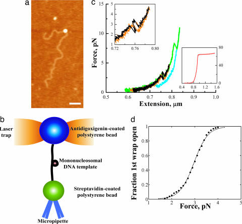
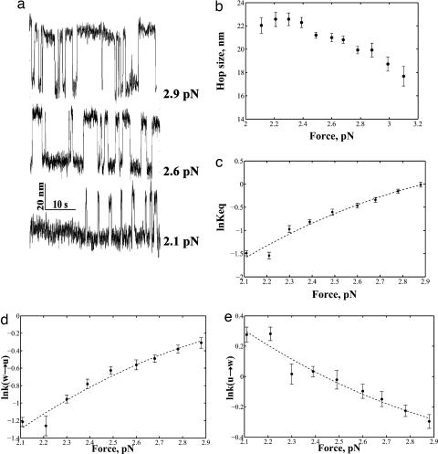
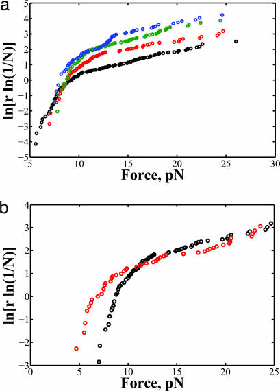
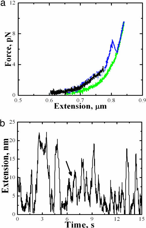
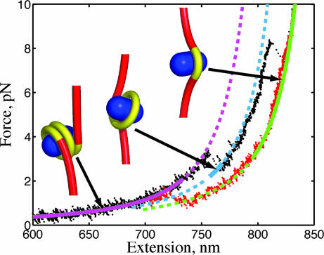
Using single-molecule optical-trapping techniques, we examined the force-induced dynamic behavior of a single nucleosome core particle. Our experiments using the DNA construct containing the 601 nucleosome-positioning sequence revealed that the nucleosome unravels in at least two major stages. The first stage, which we attributed to the unraveling of the first DNA wrap around the histone octamer, could be mechanically induced in a reversible manner, and when kept at constant force within a critical force range, exhibited two-state hopping behavior. From the hopping data, we determined the force-dependent equilibrium constant and rates for opening/closing of the outer wrap. Our investigation of the second unraveling event at various loading rates, which we attributed to the inner DNA wrap, revealed that this unraveling event cannot be described as a simple two-state process. We also looked at the behavior of the mononucleosome in a high-salt buffer, which revealed that the outer DNA wrap is more sensitive to changes in the ionic environment than the inner DNA wrap. These findings are needed to understand the energetics of nucleosome remodeling.


Conflict of interest statement

The authors declare no conflict of interest.

Figures

Fig. 1.
Fig. 1.
Pulling the mononucleosome revealed two distinct rips. (a) Experimental setup. The antidigoxigenin-coated polystyrene bead was held in the optical trap, whereas the streptavidin-coated polystyrene bead was held onto a micropipette via suction. The mononucleosome is tethered between the beads. (b) AFM image of a mononucleosome reconstituted on a 3,547-bp piece of DNA. (Scale bar, 200 nm.) (c) Representative force-extension curves of the mononucleosome. Black and green indicate when the fiber was being pulled; orange and blue indicate when the fiber was being relaxed. (Upper Left Inset) Close-up of the low-force transition. (Lower Right Inset) A typical curve for naked B-form DNA. (d) Probability of the first wrap unraveling as a function of force was obtained by summing a normalized histogram of the first wrap opened vs. force. The solid line is the probability p(E) of a two-state system of the general form: p(E) = 1/(1 + eE/kBT). Best fit of the data yielded a ΔG(unwrap) of ≈24.7 kJ/mol.
Fig. 2.
Fig. 2.
Two-state hopping of the low-force transition. (a) Length vs. time traces of the low-force transition at various forces. (b) Plots of the size of the hopping transition as a function of force. (c) Logarithm of the Keq plotted as a function of force. (d) Logarithm of the rate of going from the wrapped to the unwrapped state plotted as a function of force. (e) Logarithm of the rate of going from the unwrapped to the wrapped state as a function of force (error bar shows standard error).
Fig. 3.
Fig. 3.
Behavior of the high-force transition. (a) The force distribution of the inner DNA wrap unraveling at loading rates, r, 2.4 (black), 5.3 (red), 7.8 (green), and 11 pN/s (blue). (b) Comparison of the force distribution at different ionic strengths at r = 5.3 pN/s. The high ionic strength buffer is indicated in red.
Fig. 4.
Fig. 4.
Effect of high salt on the mononucleosome. (a) Typical force-extension curves of the mononucleosome in high ionic strength buffer. (b) Length vs. time trace of the low-force transition at 2.7 pN shows intermediates (indicated by arrow).
Fig. 5.
Fig. 5.
Force-extension curve of a spool model for the nucleosome. Theoretical curves for 1.5 wrap (purple), 1 wrap (blue), and 0.5 wrap (green) are plotted with experimental data for increasing (black) and decreasing (red) tension. The solid curve segment identifies that the state is thermodynamically preferred (minimum free energy), and the dashed segment indicates the state is stable but not preferred.

References

    1. Wolffe AP, Hayes JJ. Nucleic Acids Res. 1999;27:711–720. - PMC - PubMed
    1. Peterson CL. FEBS Lett. 2000;476:68–72. - PubMed
    1. Flaus A, Owen-Hughes T. Biopolymers. 2003;68:563–578. - PubMed
    1. Anderson JD, Widom J. J Mol Biol. 2000;296:979–987. - PubMed
    1. Li G, Levitus M, Bustamante C, Widom J. Nat Struct Mol Biol. 2005;12:46–53. - PubMed

Publication types

LinkOut - more resources