. 2006 Oct 24;103(43):16021-6.
doi: 10.1073/pnas.0607423103. Epub 2006 Oct 16.

Wild-type microglia extend survival in PU.1 knockout mice with familial amyotrophic lateral sclerosis

Affiliations

Wild-type microglia extend survival in PU.1 knockout mice with familial amyotrophic lateral sclerosis

David R Beers et al. Proc Natl Acad Sci U S A. .

Abstract

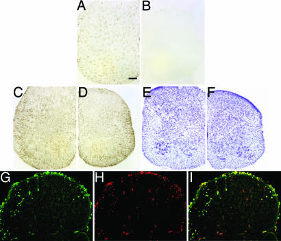
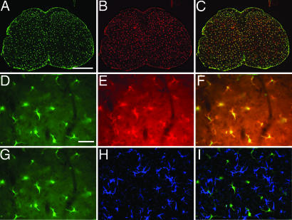
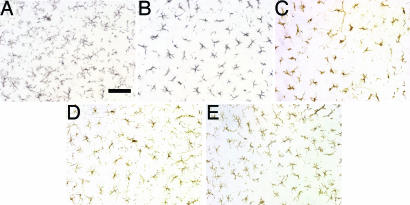
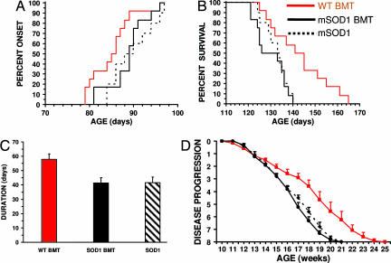
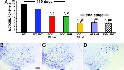
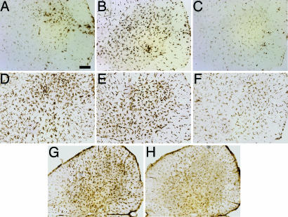
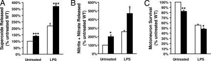
The most common inherited form of amyotrophic lateral sclerosis (ALS), a neurodegenerative disease affecting adult motoneurons, is caused by dominant mutations in the ubiquitously expressed Cu(2+)/Zn(2+) superoxide dismutase (SOD1). Recent studies suggest that glia may contribute to motoneuron injury in animal models of familial ALS. To determine whether the expression of mutant SOD1 (mSOD1(G93A)) in CNS microglia contributes to motoneuron injury, PU.1(-/-) mice that are unable to develop myeloid and lymphoid cells received bone marrow transplants resulting in donor-derived microglia. Donor-derived microglia from mice overexpressing mSOD1(G93A), an animal model of familial ALS, transplanted into PU.1(-/-) mice could not induce weakness, motoneuron injury, or an ALS-like disease. To determine whether expression of mSOD1(G93A) in motoneurons and astroglia, as well as microglia, was required to produce motoneuron disease, PU.1(-/-) mice were bred with mSOD1(G93A) mice. In mSOD1(G93A)/PU.1(-/-) mice, wild-type donor-derived microglia slowed motoneuron loss and prolonged disease duration and survival when compared with mice receiving mSOD1(G93A) expressing cells or mSOD1(G93A) mice. In vitro studies confirmed that wild-type microglia were less neurotoxic than similarly cultured mSOD1(G93A) microglia. Compared with wild-type microglia, mSOD1(G93A) microglia produced and released more superoxide and nitrite+nitrate, and induced more neuronal death. These data demonstrate that the expression of mSOD1(G93A) results in activated and neurotoxic microglia, and suggests that the lack of mSOD1(G93A) expression in microglia may contribute to motoneuron protection. This study confirms the importance of microglia as a double-edged sword, and focuses on the importance of targeting microglia to minimize cytotoxicity and maximize neuroprotection in neurodegenerative diseases.


Conflict of interest statement

The authors declare no conflict of interest.

Figures

Fig. 1.
Fig. 1.
Identification and characterization of donor-derived microglia in spinal cord sections from neonatal PU.1−/− mice. Presence (A) and absence (B) of CD11b signal on microglia in PU.1+/− mouse and PU.1−/− mouse without BMT, respectively. GFAP immunohistochemical and cresyl violet staining did not reveal any differences in astrocyte numbers or morphology, and motoneuron numbers or morphology, in spinal cord sections of 11-day-old PU.1+/− (C and E) and PU.1−/− (D and F) mice that did not receive a BMT. Green GFP (G) and red CD11b (H) signals from an 11-day-old PU.1−/− mouse. (I) Merged image of G and H. (Scale bar, 100 μm.)
Fig. 2.
Fig. 2.
Identification and characterization of donor-derived microglia in spinal cord sections from adult PU.1−/− mice. Green GFP (A) and red CD11b (B) signals from a 40-day-old PU.1−/− mouse. (C) Merged image of A and B. Higher magnification of the GFP (D) and red CD11b (E) signals from a 40-day-old PU.1−/− mouse. (F) Merged image of D and E. (G) Same image as D of the green GFP signal in spinal cord sections from 40-day-old PU.1−/− mice that received BMT from GFP donors. (H) Blue GFAP immunofluorescence indicating the presence of astrocytes. (I) Merged image of G and H demonstrating that the blue GFAP signal does not overlap the green GFP signal. (Scale bars, 100 μm in AC and 500 μm in DI.)
Fig. 3.
Fig. 3.
Adult microglia in adult PU.1 mice. CD11b positive microglia in a spinal section from 40-day-old PU.1+/− (A) and PU.1−/− (B) mice, and 140- (C), 370- (D), and 570-day-old (E) PU.1−/− mice after BMT. (Scale bar, 100 μm.)
Fig. 4.
Fig. 4.
WT BMT prolongs survival and duration in mSOD1G93A/PU.1−/− mice. (A) The onset times of mSOD1G93A mice (n = 10), and mSOD1G93A/PU.1−/− mice transplanted with WT (n = 12), or mSOD1G93A (n = 12), bone marrow were not different. (B) WT bone marrow significantly prolongs survival in mSOD1G93A/PU.1−/− mice. (C) Disease duration was 40% longer in mSOD1G93A/PU.1−/− mice receiving WT BMT. (D) Disease progression was significantly delayed in mSOD1G93A/PU.1−/− mice receiving WT BMT. Significance was evaluated by using a log rank sum test (A and B), Student's t test (C), and ANOVA with repeated measures (D).
Fig. 5.
Fig. 5.
Increased motoneuron numbers after WT BMT. (A) Motoneuron counting. n = 3 for each group; ∗, statistically different from WT mice; #, statistically different from mSOD1G93A/PU.1−/− mice with WT BMT at 110 days of age; ##, statistically different from either mSOD1G93A/PU.1+/− mice or mSOD1G93A/PU.1−/− mice with mSOD1G93A BMT at 110 days of age. (BD) Cresyl violet staining of a spinal cord section from a 110-day-old mSOD1G93A/PU.1+/− mouse (B), a 110-day-old mSOD1G93A/PU.1−/− mouse with mSOD1G93A BMT (C), and a 110-day-old mSOD1G93A/PU.1−/− mouse with WT BMT (D). Error bars indicate standard error of the mean. Significance was evaluated by using a one-way ANOVA (A). (Scale bar, 100 μm in BD.)
Fig. 6.
Fig. 6.
Characterization of microglia in vivo. (AH) Immunohistochemical staining of lumbar spinal cord from 110-day-old mice. CD68 signal from microglia/macrophages of mSOD1G93A/PU.1+/− (A), and mSOD1G93A/PU.1−/− with either mSOD1G93A BMT (B) or WT BMT (C) mice. CD11b signal from microglia/macrophages of mSOD1G93A/PU.1+/− (D), and mSOD1G93A/PU.1−/− with either mSOD1G93A BMT (E) or WT BMT (F) mice. CD40 signal from mSOD1G93A/PU.1−/− with either mSOD1G93A (G) or WT BMT (H). (Scale bar, 100 μm.)
Fig. 7.
Fig. 7.
Characterization of microglia in vitro. Open bar, WT microglia; filled bar, mSOD1G93A microglia. Superoxide (A) or nitrite+nitrate (B) released in cultures of primary microglia before and after treatment with LPS. (C) Survival of motoneuron when cocultured with microglia before and after treatment with LPS. Significance was evaluated by using an ANOVA. Error bars indicate standard error of the mean. ∗, P < 0.05; ∗∗, P < 0.01; ∗∗∗, P < 0.005.

References

    1. Cleveland DW, Rothstein JD. Nat Rev Neurosci. 2001;2:806–819. - PubMed
    1. Rosen DR, Siddique T, Patterson D, Figlewicz DA, Sapp P, Hentati A, Donaldson D, Goto J, O'Regan JP, Deng HX, et al. Nature. 1993;362:59–62. - PubMed
    1. Gurney ME, Pu H, Chiu AY, Dal Canto MC, Polchow CY, Alexander DD, Caliendo J, Hentati A, Kwon YW, Deng HX, et al. Science. 1994;264:1772–1775. - PubMed
    1. Wong PC, Pardo CA, Borchelt DR, Lee MK, Copeland NG, Jenkins NA, Sisodia SS, Cleveland DW, Price DL. Neuron. 1995;14:1105–1116. - PubMed
    1. Howland DS, Liu J, She Y, Goad B, Maragakis NJ, Kim B, Erickson J, Kulik J, DeVito L, Psaltis G, et al. Proc Natl Acad Sci USA. 2002;99:1604–1609. - PMC - PubMed

Publication types

MeSH terms