. 2007 Jan;87(1):40-8.
doi: 10.1038/labinvest.3700480. Epub 2006 Oct 16.

Comparison of lymphoblast mitochondria from normal subjects and patients with Barth syndrome using electron microscopic tomography

Affiliations

Comparison of lymphoblast mitochondria from normal subjects and patients with Barth syndrome using electron microscopic tomography

Devrim Acehan et al. Lab Invest. 2007 Jan.

Abstract

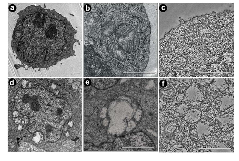
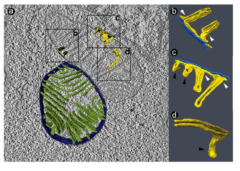
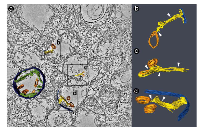
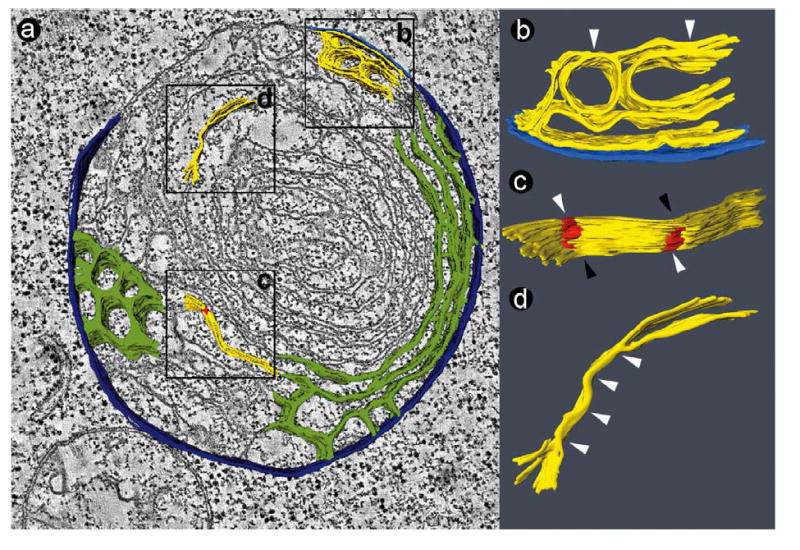
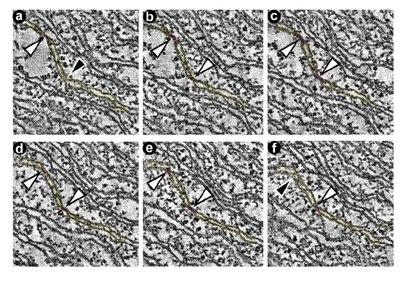
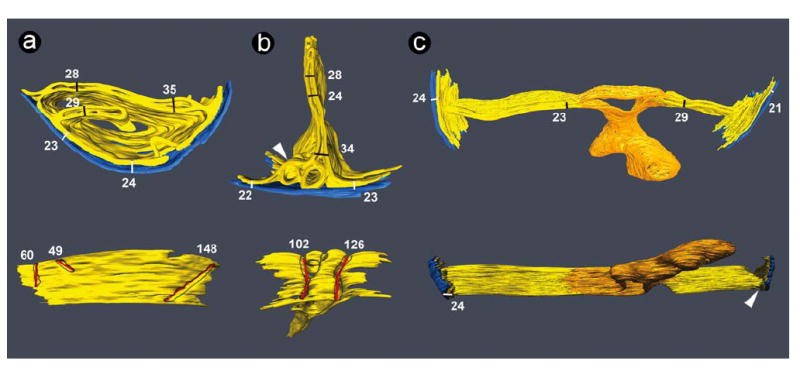
Barth syndrome (BTHS) is a mitochondrial disorder that is caused by mutations in the tafazzin gene, which affects phospholipid composition. To determine whether this defect leads to alterations in the internal three-dimensional organization of mitochondrial membranes, we applied electron microscopic tomography to lymphoblast mitochondria from BTHS patients and controls. Tomograms were formed from 50 and 150 nm sections of chemically fixed lymphoblasts and the data were used to manually segment volumes of relevant structural details. Normal lymphoblast mitochondria contained well-aligned, lamellar cristae with slot-like junctions to the inner boundary membrane. In BTHS, mitochondrial size was more variable and the total mitochondrial volume per cell increased mainly due to clusters of fragmented mitochondria inside nuclear invaginations. However, mitochondria showed reduced cristae density, less cristae alignment, and inhomogeneous cristae distribution. Three-dimensional reconstruction of BTHS mitochondria revealed zones of adhesion of the opposing inner membranes, causing obliteration of the intracrista space. We found small isolated patches of adhesion as well as extended adhesion zones, resulting in sheets of collapsed cristae packaged in multiple concentric layers. We also found large tubular structures (diameter 30-150 nm) that appeared to be derivatives of the adhesion zones. The data suggest that mitochondrial abnormalities of BTHS involve adhesions of inner mitochondrial membranes with subsequent collapse of the intracristae space.


Figures

Figure 1
Figure 1
Mitochondria in lymphoblasts from normal individuals (a-c) and BTHS patients (d-f). a: Electron micrograph of a normal lymphoblast. b: Electron micrograph of a cluster of normal mitochondria. c: Slice of a tomogram showing several normal mitochondria. d: Electron micrograph of a BTHS lymphoblast with degenerated mitochondria of variable size. e: Electron micrograph showing a giant mitochondrion inside the nuclear invagination of a BTHS lymphoblast. f: Slice of a tomogram showing fragmented mitochondria of a BTHS lymphoblast. Bars: 1 μm.
Figure 2
Figure 2
Tomogram of two normal lymphoblast mitochondria (50 nm section). a: Slice of the tomogram with surface-rendered depiction of a 3-D model, showing the peripheral compartment (outer membrane + intermembrane space + inner boundary membrane) in dark blue and the cristae in green. The boxes (0.4 μm × 0.4 μm) mark three segmented details, in which the outer membrane is shown in bright blue and the inner membrane in yellow. b, c, d: 3-D models of membranes in the corresponding boxes. The images were magnified and rotated to reveal details of interest. White arrowheads point to junctions between lamellar cristae and the inner boundary membrane. Black arrowheads point to tubular cristae.
Figure 3
Figure 3
Tomogram of a cluster of fragmented mitochondria in a BTHS lymphoblast (50 nm section). a: Slice of the tomogram with surface-rendered depiction of a 3-D model, showing the peripheral compartment (outer membrane + intermembrane space + inner boundary membrane) in dark blue, cristae in green, and large tubular structures in orange. The boxes (0.4 μm × 0.4 μm) mark three segmented details, in which the outer membrane is shown in bright blue, the inner membrane in yellow, and the tubular structures in orange. b, c, d: 3-D models of membranes in the corresponding boxes. The images were magnified and rotated to reveal details of interest. White arrowheads point to zones of inner membrane adhesion. The large tubes (orange) are connected to collapsed cristae membranes.
Figure 4
Figure 4
Tomogram of a giant mitochondrion in a BTHS lymphoblast (50 nm section). a: Slice of the tomogram with surface-rendered depiction of a 3-D model, showing the peripheral compartment (outer membrane + intermembrane space + inner boundary membrane) in dark blue and the cristae in green. Two types of cristae arrangement are visible: a honeycomb pattern and a concentric pattern. The boxes (0.4 μm × 0.4 μm) mark three segmented details, in which the outer membrane is shown in bright blue and the inner membrane in yellow. b, c, d: 3-D models of membranes in the corresponding boxes. The images were magnified and rotated to reveal details of interest. White arrowheads point to zones of inner membrane adhesion. In Figure 4c, the red zones mark adhesions of the inner membranes, white arrowheads point to areas with collapsed intracrista space, and black arrowheads point to areas with open intracrista space (see also Figure 5).
Figure 5
Figure 5
Representative slices through the tomogram showing segmentations used to create the model in Figure 4c. a-f: The series of slices demonstrates the presence of adhesion zones between opposing inner membranes (white arrowheads). Adhesion zones alternate with zones that have an open intermembrane space (black arrowheads).
Figure 6
Figure 6
3-D models of tomograms obtained from 150 nm sections. The outer membranes are shown in blue and the inner membranes are shown in yellow. Each model is depicted in two views rotated approximately 90° relative to each other. Numbers represent dimensions of the corresponding bars in nm. White bars show the width of the mitochondrial periphery (outer membrane + intermembrane space + inner boundary membrane), black bars show the cristae width, and red bars show the length of cristae junctions at the level of the inner boundary membrane. a: Mitochondrial membranes from a normal lymphoblast. In the lower image, the outer membrane was deleted to reveal the openings of three cristae junctions, outlined in grey. The left crista junction (60 nm) extends beyond the segmented volume. Dimensions in the upper image are consistent with previously published data (21, 27, 29, 30). b: Mitochondrial membranes from a BTHS lymphoblast. In the lower image, the outer membrane was deleted to reveal the openings of two cristae junctions, outlined in grey. The arrowhead points to a region of interconnected cristae with honeycomb pattern. c: Mitochondrial membranes from a BTHS lymphoblast. Cristae are connected to a membrane (orange) that encloses a large cavity with multiple branches. This structure is specific to BTHS mitochondria and is also seen in Figure 3 (orange tubes). The arrowhead points to the lateral view of a crista junction.

References

    1. Barth PG, Scholte HR, Berden JA, et al. An X-linked mitochondrial disease affecting cardiac muscle, skeletal muscle and neutrophil leukocytes. J Neurol Sci. 1983;62:327–355. - PubMed
    1. Kelley RI, Cheatham JP, Clark BJ, et al. X-linked dilated cardiomyopathy with neutropenia, growth retardation, and 3-methylglutaconic aciduria. J Pediatr. 1991;119:738–747. - PubMed
    1. Barth PG, Wanders RJA, Vreken P, et al. X-linked cardioskeletal myopathy and neutropenia (Barth syndrome) (MIM 302060) J Inher Metab Dis. 1999;22:555–567. - PubMed
    1. Bione S, D’Adamo P, Maestrini E, et al. A novel X-linked gene, G4.5. is responsible for Barth syndrome. Nature Genetics. 1996;12:385–389. - PubMed
    1. Ma L, Vaz FM, Gu Z, et al. The human TAZ gene complements mitochondrial dysfunction in the yeast taz1Δ mutant. Implications for Barth syndrome. J Biol Chem. 2004;279:44394–44399. - PubMed

Publication types

MeSH terms