. 2007 Jan;25(1):163-9.
doi: 10.1016/j.nbd.2006.09.002. Epub 2006 Oct 10.

Brain injury impairs dentate gyrus inhibitory efficacy

Affiliations

Brain injury impairs dentate gyrus inhibitory efficacy

David P Bonislawski et al. Neurobiol Dis. 2007 Jan.

Abstract

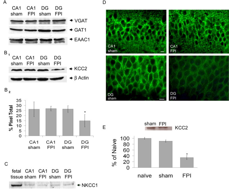
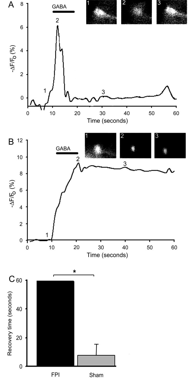
Every 23 s, a person sustains a traumatic brain injury in the United States leaving many patients with substantial cognitive impairment and epilepsy. Injury-induced alterations in the hippocampus underpin many of these disturbances of neurological function. Abnormalities in the dentate gyrus are likely to play a major role in the observed pathophysiology because this subregion functions as a filter impeding excessive or aberrant activity from propagating further into the circuit and following experimental brain injury, the dentate gyrus becomes more excitable. Although alteration in excitation or inhibition could mediate this effect in the dentate gyrus, we show a key role played by an impairment of GABA(A)ergic inhibition. The efficacy of GABA(A)-mediated inhibition depends on a low [Cl-]i that is maintained by neuronal K-Cl co-transporter 2 (KCC2). Using fluid percussion injury (FPI) in the mouse, we demonstrate significant reductions in KCC2 protein and mRNA expression in the dentate gyrus that causes a depolarizing shift in GABA(A) reversal potential, due to impaired chloride clearance, resulting in reduced inhibitory efficiency. This study elucidates a novel mechanism underlying diminished dentate gyrus inhibitory efficacy and provides an innovative target for the development of potential therapeutics to restore the severe pathological consequences of traumatic brain injury.


Figures

Figure 1
Figure 1. KCC2 protein and mRNA expression is significantly reduced in dentate gyrus seven days following FPI
A) Representative Western blot showing no difference in expression of VGAT (55 kDa), GAT1 (67 kDa) and EAAC1 (69 kDa) within isolated CA1 and dentate gyrus (DG) from ipsilateral (injured) hippocampus of both sham and FPI animals (B1) Representative Western blot showing expression of KCC2 (142 kDa) within isolated CA1 and dentate gyrus from ipsilateral (injured) hippocampus of both sham and FPI animals. Control for protein loading is shown for β-actin (45kDa). (B2) Densitometry histogram demonstrating significant reduced expression of KCC2 in the dentate gyrus of injured animals. (C) Representative western blot showing expression of NKCC1 (145kDa) within isolated CA1 and DG from ipsilateral (injured) hippocampus of both sham and FPI animals compared to fetal brain tissue. (D) KCC2 immunohistochemistry in area CA1 (top panel) and dentate gyrus (lower panel) 7 days following FPI. Scale bar is 10 μm. (E) Inset: Representative phosphoimage of total mRNA isolated from DG tissue from FPI and sham mice. Histogram: Real time RT-PCR analysis of KCC2 mRNA expression in dentate gyrus 7 days following FPI compared to sham and naive.
Figure 2
Figure 2. Brain injury induces a depolarized shift in GABA reversal potential in dentate gyrus
(A) A representative IV curve demonstrating EGABA to be −73 mV recorded in a dentate gyrus neuron from a sham animal. As a control the perforated patch was ruptured, thereby entering into the whole cell configuration and EGABA shifts to approximately 0. Insets are current recordings in response to the displayed voltage ramp protocol in the absence (left) and presence (right) of focally applied agonist (GABA). (B) Representative current-voltage curve shows a significant shift in EGABA in a dentate gyrus neuron recorded in a slice from an FPI animal to −51 mV. When the superfusing aCSF is changed to low K+ (1 mM, 2–3 min) aCSF the curve shifts toward EGABA observed in sham animals. Inset: negative shift in EGABA in 3 DG neurons recorded in slices from injured animals when aCSF is switched to low K+. (C) Histogram of the hyperpolarizing response to focally applied GABA (100μM) recorded in current clamp mode demonstrating a significant decrease in the amplitude of the response recorded in dentate gyrus from injured animals.
Figure 3
Figure 3. Intracellular chloride homeostasis is disrupted seven days post FPI
(A) In a dentate granule neuron from a sham animal, GABA (bath application 30μM, 10 sec) caused an influx of chloride, measured by MEQ fluorescence, which was quickly cleared. Inset: MEQ fluorescent image of same dentate gyrus neuron at: 1) baseline 2) immediately after GABA application 3) 20 seconds after GABA application. (B) In a dentate granule neuron from an FPI animal, GABA application caused an influx of chloride, however clearance was significantly protracted to > 60 seconds. inset: image of same dentate granule neuron at: 1) baseline 2) immediately after GABA application 3) 30 seconds after GABA application. (C) Histogram of group data summarizing the time for fluorescence intensity to return to baseline following application of GABA.

References

    1. Annegers JF, Hauser WA, Coan SP, Rocca WA. A population-based study of seizures after traumatic brain injuries. N Engl J Med. 1998;338:20–24. - PubMed
    1. Asikainen I, Kaste M, Sarna S. Early and late posttraumatic seizures in traumatic brain injury rehabilitation patients: brain injury factors causing late seizures and influence of seizures on long-term outcome. Epilepsia. 1999;40:584–589. - PubMed
    1. BIAUSA. TBI Statistics. 2004.
    1. Buzsaki G, Leung LW, Vanderwolf CH. Cellular bases of hippocampal EEG in the behaving rat. Brain Res. 1983;287:139–171. - PubMed
    1. Cave CB, Squire LR. Equivalent impairment of spatial and nonspatial memory following damage to the human hippocampus. Hippocampus. 1991;1:329–340. - PubMed

Publication types

MeSH terms