. 2006 Oct 19;52(2):255-69.
doi: 10.1016/j.neuron.2006.09.037.

Brain-specific phosphorylation of MeCP2 regulates activity-dependent Bdnf transcription, dendritic growth, and spine maturation

Affiliations

Brain-specific phosphorylation of MeCP2 regulates activity-dependent Bdnf transcription, dendritic growth, and spine maturation

Zhaolan Zhou et al. Neuron. .

Abstract

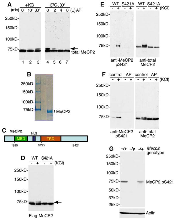
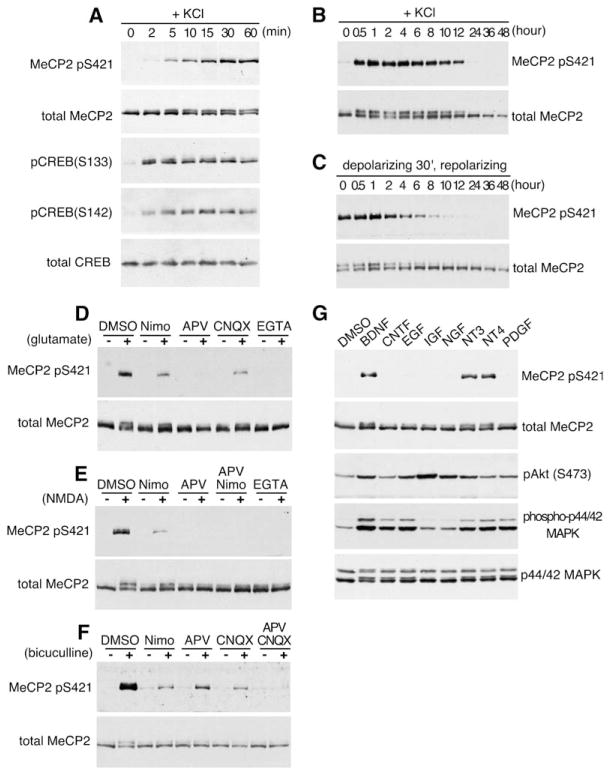
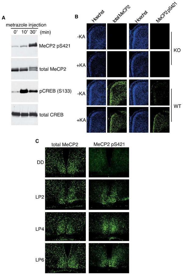
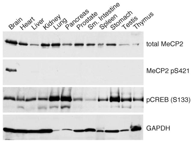
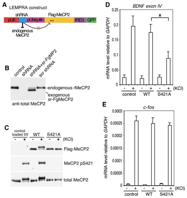
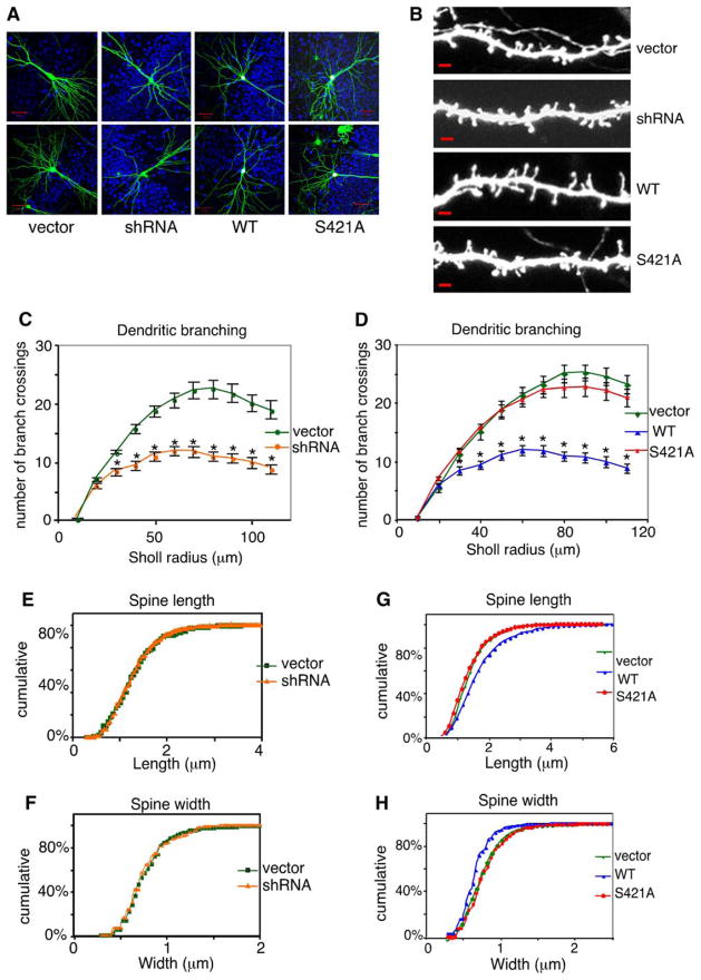
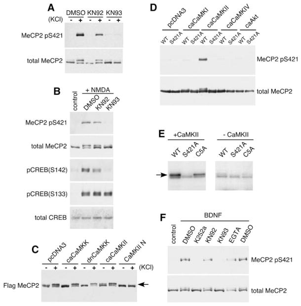
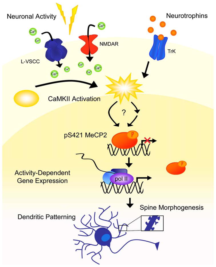
Mutations or duplications in MECP2 cause Rett and Rett-like syndromes, neurodevelopmental disorders characterized by mental retardation, motor dysfunction, and autistic behaviors. MeCP2 is expressed in many mammalian tissues and functions as a global repressor of transcription; however, the molecular mechanisms by which MeCP2 dysfunction leads to the neural-specific phenotypes of RTT remain poorly understood. Here, we show that neuronal activity and subsequent calcium influx trigger the de novo phosphorylation of MeCP2 at serine 421 (S421) by a CaMKII-dependent mechanism. MeCP2 S421 phosphorylation is induced selectively in the brain in response to physiological stimuli. Significantly, we find that S421 phosphorylation controls the ability of MeCP2 to regulate dendritic patterning, spine morphogenesis, and the activity-dependent induction of Bdnf transcription. These findings suggest that, by triggering MeCP2 phosphorylation, neuronal activity regulates a program of gene expression that mediates nervous system maturation and that disruption of this process in individuals with mutations in MeCP2 may underlie the neural-specific pathology of RTT.


Figures

Figure 1
Figure 1. Identification of MeCP2 S421 as a Site of Membrane Depolarization-Induced Phosphorylation
(A) Antitotal MeCP2 western blots of whole-cell lysates (lanes 1–3) or nuclear extracts treated with alkaline phosphatase (lanes 5– 7) prepared from E18 + 5 DIV dissociated rat cortical neurons harvested at the indicated times following 55 mM KCl stimulation. The arrow indicates the slow-migrating form of MeCP2 induced in response to membrane depolarization. AP: Nuclear extracts were treated with 2, 4, or 8 U of calf intestinal alkaline phosphatase for 30 min at 37°C prior to SDS-PAGE separation. Note: MeCP2 runs at approximately 75 kDa on an 8% SDS-PAGE gel. All protein samples in this study were separated on an 8% gel unless otherwise noted. (B) Coomassie blue staining of MeCP2 protein purified from rat brain nuclear extract. Both bands were excised and subjected to tandem MS/MS sequencing for identification of phosphorylation sites. (C) A schematic of MeCP2 illustrating the positions of phosphorylation sites relative to the methyl-CpG-binding domain (MBD), nuclear localization signal (NLS), and the transcriptional repression domain (TRD). (D) Identification of S421 as a residue required for membrane depolarization-induced phosphorylation of MeCP2. FLAG-tagged wild-type or S421A MeCP2 was transfected into E18 + 2 DIV cortical neurons. Two days later, extracts were prepared from untreated neurons or neurons membrane depolarized for 60 min and subjected to western blotting with the anti-FLAG antibody. (E) A phospho specific antibody detects phosphorylation of MeCP2 at S421. FLAG immunoprecipitates from E18 + 5 DIV cortical neurons transfected with FLAG-tagged wild-type or S421A MeCP2 were immunoblotted with antitotal MeCP2 or anti-MeCP2 pS421 antibodies. The anti-MeCP2 pS421 antibody recognizes a band at a position corresponding to that of the slow-migrating species of MeCP2. (F) Membrane depolarization triggers the phosphorylation of MeCP2 at S421. Antitotal MeCP2 and anti-MeCP2 pS421 western blots of nuclear extracts prepared from unstimulated or membrane-depolarized E18 + 5 DIV cortical neurons, either untreated (control) or incubated with alkaline phosphatase for 30 min at 37°C (AP). Unlike other experiments, cultures were not treated with TTX prior to stimulation in this experiment. (G) The anti-MeCP2 pS421 antibody specifically recognizes MeCP2. Brain lysates from C57BL/6 wild-type Mecp2+/+, heterozygous Mecp2/+, or Mecp2/y null mice were probed with anti-MeCP2 pS421 or anti-actin antibodies.
Figure 2
Figure 2. Characterization of S421 as a Site of Activity-Dependent Phosphorylation on MeCP2
(A–C) Kinetics of MeCP2 S421 phosphorylation and dephosphorylation. Lysates were prepared from rat E18 + 5 DIV cortical neurons membrane depolarized for the indicated times (A and B) or membrane depolarized for 30 min and repolarized with culture medium for the indicated times (C). (D–F) MeCP2 is phosphorylated at S421 in response to glutamate (D), NMDA (E), and bicuculline (F) in a calcium-dependent manner. Antitotal MeCP2 and anti-MeCP2 pS421 western blots of extracts from E18 + 12 DIV hippocampal neurons (D and E) or P1 + 12 DIV hippocampal neurons (F) treated for 1 hr with the indicated stimuli after a 1 hr pretreatment with the indicated blockers. Nimo, nimodipine. (G) MeCP2 S421 phosphorylation is triggered by neurotrophins. Western blot analysis of whole-cell extracts prepared from E18 + 12 DIV hippocampal neurons stimulated for 1 hr with BDNF, CNTF, EGF, IGF, NGF, NT3, NT4, or PDGF.
Figure 3
Figure 3. Phosphorylation of MeCP2 at S421 Is Triggered by Synaptic Activity in the Intact Brain
(A) Whole-cell lysates were prepared from P19 rat brains 0, 10, or 30 min after metrazole-based seizure induction and incubated with antibodies specific to MeCP2 pS421, total MeCP2, pCREB (S133), or total CREB. (B) Fluorescent immunohistochemistry of coronal cryosections from mouse parietal cortex collected from wild-type (WT) or Mecp2/y (KO) mice 3 hr after kainic acid-based seizure induction using antibodies specific for MeCP2 pS421 or total MeCP2. Sections were counterstained with the nuclear dye Hoechst 33342. (C) Light exposure during subjective night induces the phosphorylation of MeCP2 S421 in the suprachiasmatic nucleus (SCN). Wild-type mice were entrained to a 12hr:12hr light-dark cycle and then transferred to constant darkness. Two days later, entrained mice were either maintained in darkness (DD) or exposed to light for 2, 4, or 6 hr (LP2, LP4, LP6). Adjacent cryosections were alternately immunostained with antibodies specific for MeCP2 pS421 or total MeCP2.
Figure 4
Figure 4. MeCP2 Is Phosphorylated at S421 Specifically in the Brain
Western analysis of whole-cell lysates prepared from different mammalian tissues using antibodies specific to total MeCP2, MeCP2 pS421, and pCREB (S133). Fresh tissues were collected from a 4-week-old male mouse, directly homogenized into 1.5% SDS sample buffer at a ratio of 50 mg/ml w:v, and separated on a 12% SDS-PAGE gel. The same blot was reprobed with an anti-GAPDH antibody as an indicator of sample loading.
Figure 5
Figure 5. Phosphorylation of MeCP2 at S421 Is Required for Activity-Induced Induction of Bdnf Transcription
(A) The lentivirus-mediated protein-replacement assay (LEMPRA) construct contains two expression cassettes: (1) the pU6 pol III promoter directing the expression of an shRNA directed against endogenous MeCP2 and (2) the human Ubiquitin-C promoter driving the expression of a bicistronic cassette comprised of FLAG-tagged shRNA-resistant wild-type or S421A mutant MeCP2 followed by an internal ribosomal entry site (IRES) directing GFP expression. When packaged into and delivered by a lentivirus, the construct is effective in replacing MeCP2 in greater than 90% of neurons in a culture. (B and C) LEMPRA can effectively reduce endogenous MeCP2 and express functional FLAG-tagged MeCP2 variants of choice. E18 rat hippocampal neurons were infected at 1 DIV with indicated lentiviruses. Extracts were prepared at 4 DIV (B) or 7 DIV (C) and incubated with antibodies specific to FLAG, MeCP2 pS421, or total MeCP2. shRNA, small hairpin RNA against MeCP2; sr-FgMP2, FLAG-tagged shRNA-resistant mouse MeCP2, e2 form; scr shRNA, scrambled shRNA; rMeCP2, endogenous rat MeCP2. Note that in (C), 5-fold more control lysates were loaded than wild-type or S421A-MeCP2 lysates for comparison of expression levels. (D and E) E18 + 1 DIV hippocampal neurons were infected with control, wild-type, or S421A mutant MeCP2 LEMPRA lentiviruses. Total RNA was collected at 7 DIV from infected neurons that were either left untreated (−) or membrane depolarized with 55 mM KCl for 3 hr (+). Bdnf exon IV (D) and c-fos (E) mRNA levels were measured by quantitative RT-PCR and normalized to a Gapdh control (data are mean±SEM; *p < 0.01, Bonferroni-corrected multiple comparison after ANOVA). Note that the nomenclature for the exons of the Bdnf gene has changed due to the identification of additional Bdnf transcripts in rodent; Bdnf exon IV was previously known as Bdnf exon III, for example, in Chen et al. [2003]).
Figure 6
Figure 6. Phosphorylation of MeCP2 at S421 Mediates MeCP2-Dependent Regulation of Dendritic Growth and Spine Maturation
(A) Organotypic hippocampal slices were prepared from P5–7 rat pups and biolistically transfected after 2 days in culture with LEMPRA-based plasmid DNA encoding a control vector (in which the shRNA directed against MeCP2 is replaced with a scrambled shRNA and a stop codon is introduced directly after the FLAG epitope), the anti-MeCP2 shRNA only, wild-type MeCP2, or S421A MeCP2, in combination with a dual-promoter plasmid expressing eGFP and Bcl-XL. Representative 25× images of 7 DIV transfected pyramidal neurons are shown. Scale bar, 50 μm. Pseudocoloring: green, GFP; red, anti-FLAG; blue, antitotal MeCP2. (B) Representative 63× GFP images of dendritic spines from control-, anti-MeCP2 shRNA-, wild-type MeCP2-, or S421A MeCP2-transfected neurons. Scale bar, 2 μm. (C and D) Quantification of dendritic branch complexity by Sholl analysis (data are presented as mean ± SEM, *p < 0.05, ANOVA). (E and F) shRNA-mediated knockdown of MeCP2 does not affect dendritic spine morphology (p > 0.05, K-S test) (G and H) Quantification of the effects of wild-type or S421A mutant MeCP2 overexpression on dendritic spine length (G) and width (H) (p < 0.05 for wild-type versus S421A or wild-type versus control, K-S test).
Figure 7
Figure 7. CaMKII Mediates the Phosphorylation of MeCP2 at S421
(A and B) Western blot analysis of extracts from E18 + 10 DIV hippocampal neurons that were pretreated for 60 min with the CaMKII inhibitor KN93, the inactive analog KN92, or vehicle control (DMSO), followed by stimulation with 55 mM KCl (A) or 20 μM NMDA (B) for 30 min. (C) E18 + 5 DIV cortical neurons were cotransfected with FLAG-tagged MeCP2 and vector control, ca-CaMKK, dn-CaMKK, ca-CaMKII, or CaMKII-N. Extracts were prepared from these neurons left unstimulated (−) or membrane depolarized for 1 hr (+) 2 days after transfection and probed with antibodies specific to the FLAG epitope. ca, constitutive-active; dn, dominant-negative. (D) Western blot analysis using anti-MeCP2 pS421 or anti-FLAG antibodies of extracts from HEK293T cells cotransfected with FLAG-MeCP2 and vector control, ca-CaMKI, ca-CaMKII, ca-CaMKIV, or ca-Akt. (E) CaMKII phosphorylates MeCP2 in vitro. FLAG-tagged wild-type, S421A mutant MeCP2, or C5A mutant MeCP2 (S341A, S350A, S360A, S385A, S399A) were purified from HEK293T cells, coincubated with recombinant constitutively active CaMKII (left panel) and 32P-ATP, and separated on an SDS-PAGE gel. The right panel shows the results of a control reaction in the absence of any added kinase. (F) BDNF-dependent induction of MeCP2 S421 phosphorylation is likely mediated by CaMKII. Western blot analysis of whole-cell extracts prepared from E18 + 12 DIV hippocampal neurons that were treated with indicated agents for 60 min followed by BDNF treatment for 60 min. K252a is an inhibitor of the BDNF receptor TrkB.
Figure 8
Figure 8
Regulation of Dendritic Morphogenesis and Spine Maturation by Activity-Dependent Phosphorylation of MeCP2

References

    1. Amir RE, Van den Veyver IB, Wan M, Tran CQ, Francke U, Zoghbi HY. Rett syndrome is caused by mutations in X-linked MECP2, encoding methyl-CpG-binding protein 2. Nat Genet. 1999;23:185–188. - PubMed
    1. Ariani F, Mari F, Pescucci C, Longo I, Bruttini M, Meloni I, Hayek G, Rocchi R, Zappella M, Renieri A. Realtime quantitative PCR as a routine method for screening large rearrangements in Rett syndrome: report of one case of MECP2 deletion and one case of MECP2 duplication. Hum Mutat. 2004;24:172–177. - PubMed
    1. Asaka Y, Jugloff DG, Zhang L, Eubanks JH, Fitzsimonds RM. Hippocampal synaptic plasticity is impaired in the Mecp2-null mouse model of Rett syndrome. Neurobiol Dis. 2006;21:217–227. - PubMed
    1. Ballas N, Grunseich C, Lu DD, Speh JC, Mandel G. REST and its corepressors mediate plasticity of neuronal gene chromatin throughout neurogenesis. Cell. 2005;121:645–657. - PubMed
    1. Bienvenu T, Chelly J. Molecular genetics of Rett syndrome: when DNA methylation goes unrecognized. Nat Rev Genet. 2006;7:415–426. - PubMed

Publication types

MeSH terms

Substances