. 2006 Oct 19;52(2):271-80.
doi: 10.1016/j.neuron.2006.07.023.

Definition of estrogen receptor pathway critical for estrogen positive feedback to gonadotropin-releasing hormone neurons and fertility

Affiliations

Definition of estrogen receptor pathway critical for estrogen positive feedback to gonadotropin-releasing hormone neurons and fertility

Tim M Wintermantel et al. Neuron. .

Abstract

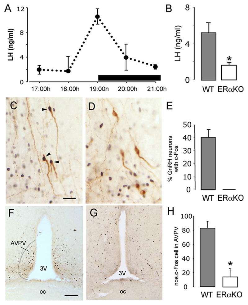
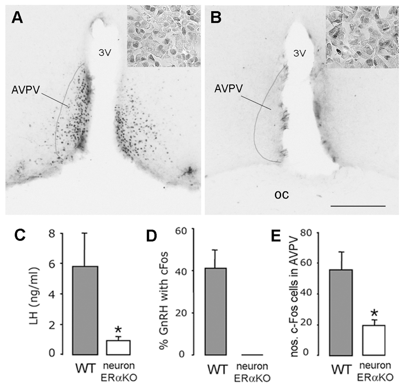
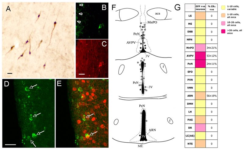
The mechanisms through which estrogen regulates gonadotropin-releasing hormone (GnRH) neurons to control mammalian ovulation are unknown. We found that estrogen positive feedback to generate the preovulatory gonadotropin surge was normal in estrogen receptor beta knockout (ERbeta) mutant mice, but absent in ERalpha mutant mice. An ERalpha-selective compound was sufficient to generate positive feedback in wild-type mice. As GnRH neurons do not express ERalpha, estrogen positive feedback upon GnRH neurons must be indirect in nature. To establish the cell type responsible, we generated a neuron-specific ERalpha mutant mouse line. These mice failed to exhibit estrogen positive feedback, demonstrating that neurons expressing ERalpha are critical. We then used a GnRH neuron-specific Pseudorabies virus (PRV) tracing approach to show that the ERalpha-expressing neurons innervating GnRH neurons are located within rostral periventricular regions of the hypothalamus. These studies demonstrate that ovulation is driven by estrogen actions upon ERalpha-expressing neuronal afferents to GnRH neurons.


Figures

Figure 1
Figure 1. Absence of estrogen positive feedback in ERα mutant mice.
A. Depicts the profile of the LH surge in wild-type ovariectomized mice treated with estrogen. n=4-5 at each time point. B. Mean (+SEM) LH levels in wildtype-littermates (n=7) and ERα mutant mice (n=8), ovariectomized, treated with estrogen and killed at 19:00h, * p<0.05. C-E, Dual-labelling c-Fos (black nuclei) and GnRH (brown cytoplasmic staining) immunocytochemistry in wild-type (C) and ERα mutant (D) mice. Whereas approximately 40% of GnRH neurons express c-Fos (C, arrowheads and E) in wild-type mice, no GnRH neurons express c-Fos in ERα mutant mice (D). F-H, Low-power photomicrographs showing the location of many c-Fos-expressing cells within the AVPV of wild-type mice (F) compared with few in ERα mutant mice (G). The mean (+SEM) number of c-Fos cells per unit area in the AVPV is shown in H. p<0.05. Scale bars represent 30μm in C and 100μm in F.
Figure 2
Figure 2. Positive feedback actions of estrogen appear normal in ERβ mutant mice.
A. Mean (+SEM) LH levels in wildtype-littermates (n=7) and ERβ mutant mice (n=7), ovariectomized, treated with estrogen and killed at 19:00h. B. The percentage of GnRH neurons (+SEM) found to express c-Fos in wildtype-littermates (n=7) and ERα mutant mice (n=8), ovariectomized, treated with estrogen and killed at 19:00h. C. The number of c-Fos-expressing neurons detected within the AVPV of wildtype-littermates (n=7) and ERα mutant mice (n=8), ovariectomized, treated with estrogen and killed at 19:00h.
Figure 3
Figure 3. Absence of estrogen positive feedback in neuron-specific ERα mutant mice.
A,B. show ERα expression in the rostral hypothalamus within the anteroventral periventricular nucleus (AVPV) of (A) control (ERαfl/fl) and (B) neuron-specific ERα mutant (ERαfl/fl;CamKiCre) mice. Scale bar is 100μm, 3V= third ventricle, OC = optic chiasm. Insets show equivalent ERα staining in the anterior pituitary of (A) control and (B) neuron-specific ERα mutant mice. C. Mean (+SEM) LH levels in control-littermates (n=5) and ERαfl/fl;CamKiCre mice (n=4), ovariectomized, treated with estrogen and killed at 19:00h. * p<0.05 D. Approximately 40% of GnRH neurons express c-Fos in control mice ovariectomized, treated with estrogen and killed at 19:00h whereas none are found in ERαfl/fl;CamKiCre mice. E. The number of c-Fos-expressing neurons detected within the AVPV of controls and ERαfl/fl;CamKiCre mice, ovariectomized, treated with estrogen and killed at 19:00h. * p<0.05.
Figure 4
Figure 4. Ovarian and uterine phenotype of neuron-specific ERα mutant mice.
A,B. reproductive tract of control ERαfl/fl (A) and mutant ERαfl/fl;CamKiCre (B) mice showing fluid-filled uteri in mutants. C,D H&E staining of uteri from control ERαfl/fl (C) and mutant ERαfl/fl;CamKiCre (D) mice shows atrophy and lack of glandular structures in the mutant. E,F. H&E staining of ovaries from control ERαfl/fl (E) and mutant ERαfl/fl;CamiCre (F) mice shows increased numbers of antral follicles and lack of corpora lutea in the mutant.
Figure 5
Figure 5. Identification of ERα-expressing primary afferents to GnRH neurons using GnRH-specific viral mediated tracing.
A. Restricted expression of Cre (black nuclei) in GnRH neurons (brown cytoplasm) of transgenic GnRH-Cre mice. B,C. A rostral preoptic area GnRH neuron adjacent to the injection site (arrowheads) exhibiting GFP immunoreactivity 48h following injection of Ba2001. B shows GFP immunoreactivity and C is GnRH immunostaining. D. Four neurons (arrows) in the AVPV exhibiting GFP immunoreactivity 72h after Ba2001 injection into the rostral preoptic area. E. Dual-labelling for GFP (green) and ERα (red) reveals that 2 of these cells (arrows) express ERα. F. Schematic brain maps demonstrating GFP-immunoreactive neurons (open stars) and GFP+ERα immunoreactive (filled stars) cells detected in individual 30μm-thick coronal brain sections at three levels in the hypothalamus. Abbreviations, aca, anterior commisure; ARN, arcuate nucleus; AVPV, anteroventral periventricular nucleus; f, fornix; MnPO, median preoptic nucleus; ME, median eminence, PeN, periventricular nucleus; 3V, third ventricle. Scale bar is 15μm in A,C,D. G. Summary of locations of primary afferents to GnRH neurons following stereotaxic injection of Ba2001 PRV into the rPOA of GnRH-Cre mice. Various colors in boxes adjacent to listed brain areas indicate ranges of mean numbers of GFP-positive neurons identified in female mice (n=6). Brain regions in which labelled cells were detected in <5 of the 6 mice are indicated as yellow boxes (variable). Right-most column indicates the mean percentage (±SEM) of GFP-positive neurons expressing ERα immunoreactivity. Abbreviations; ARN, arcuate nucleus; AVPV, anteroventral periventricular nucleus; DBB, diagonal band of Broca; DMH, dorsomedial hypothalamus; DR, dorsal raphe; LC, locus coereulus; LH, lateral hypothalamus; LS, lateral septum; MnPO, median preoptic nucleus; MPN, medial preoptic nucleus; MS, medial septum; NTS, nucleus tractus solitarius; PAG, periaqueductal grey; PeN, periventricular nucleus; PVN, paraventricular nucleus; SFO, subfornical organ; VMN, ventromedial nucleus.

References

    1. Abraham IM, Han K, Todman MG, Korach KS, Herbison AE. Estrogen receptor β mediates rapid estrogen actions on gonadotropin-releasing hormone neurons in vivo. J Neurosci. 2003;23:5771–5777. - PMC - PubMed
    1. Bronson FH. The regulation of luteinizing hormone secretion by estrogen: relationships among negative feedback, surge potential, and male stimulation in juvenile, peripubertal, and adult female mice. Endocrinology. 1981;108:506–516. - PubMed
    1. Burgin KE, Waxham MN, Rickling S, Westgate SA, Mobley WC, Kelly PT. In situ hybridization histochemistry of Ca2+/calmodulin-dependent protein kinase in developing rat brain. J Neurosci. 1990;10:1788–1798. - PMC - PubMed
    1. Card JP, Enquist LW. Neurovirulence of pseudorabies virus. Crit Rev Neurobiol. 1995;9:137–162. - PubMed
    1. Casanova E, Fehsenfeld S, Mantamadiotis T, Lemberger T, Greiner E, Stewart AF, Schutz G. A CamKIIalpha iCre BAC allows brain-specific gene inactivation. Genesis. 2001;31:37–42. - PubMed

Publication types

MeSH terms