

Fine-tuning of awake prefrontal cortex neurons by clozapine: comparison with haloperidol and N-desmethylclozapine
- PMID: 17046721
- PMCID: PMC2910426
- DOI: 10.1016/j.biopsych.2006.05.016
Fine-tuning of awake prefrontal cortex neurons by clozapine: comparison with haloperidol and N-desmethylclozapine
Abstract
Background: Mechanisms underlying clozapine's better clinical efficacy in schizophrenia remain poorly understood. The prefrontal cortex (PFC) has been implicated as a primary site for the therapeutic effects of clozapine; however, evidence for how clozapine influences the activity of PFC neurons in behaviorally relevant contexts is lacking.
Methods: Ensemble single unit recording in awake rats was used to measure the activity of PFC neurons in response to clozapine, its main metabolite N-desmethylclozapine (DMClz), and the typical antipsychotic drug haloperidol during baseline conditions and after treatment with the N-methyl-D-aspartate antagonist MK801. Behavioral stereotypy was scored during recording.
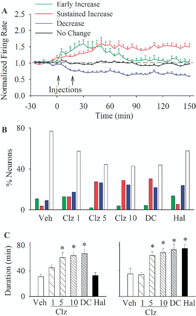
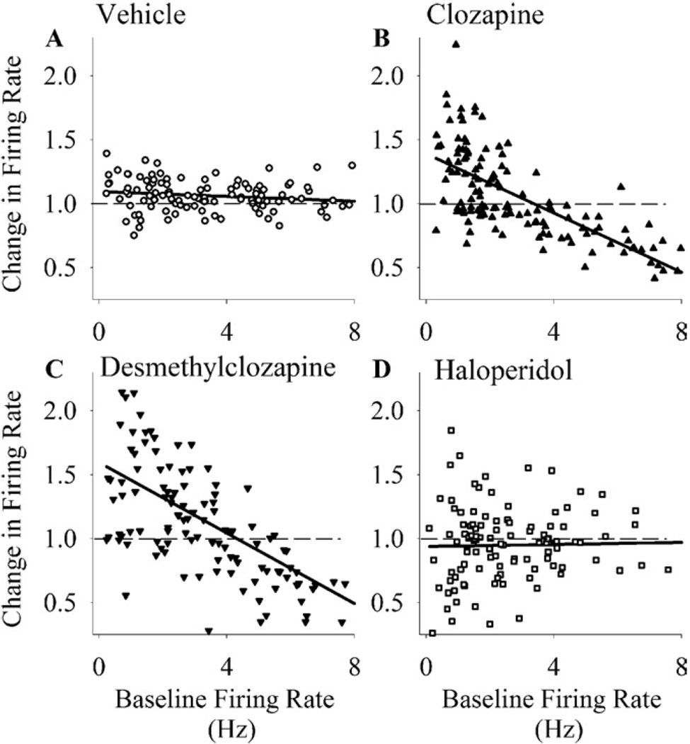
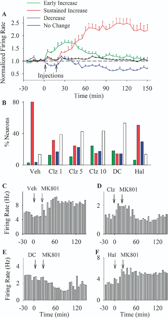
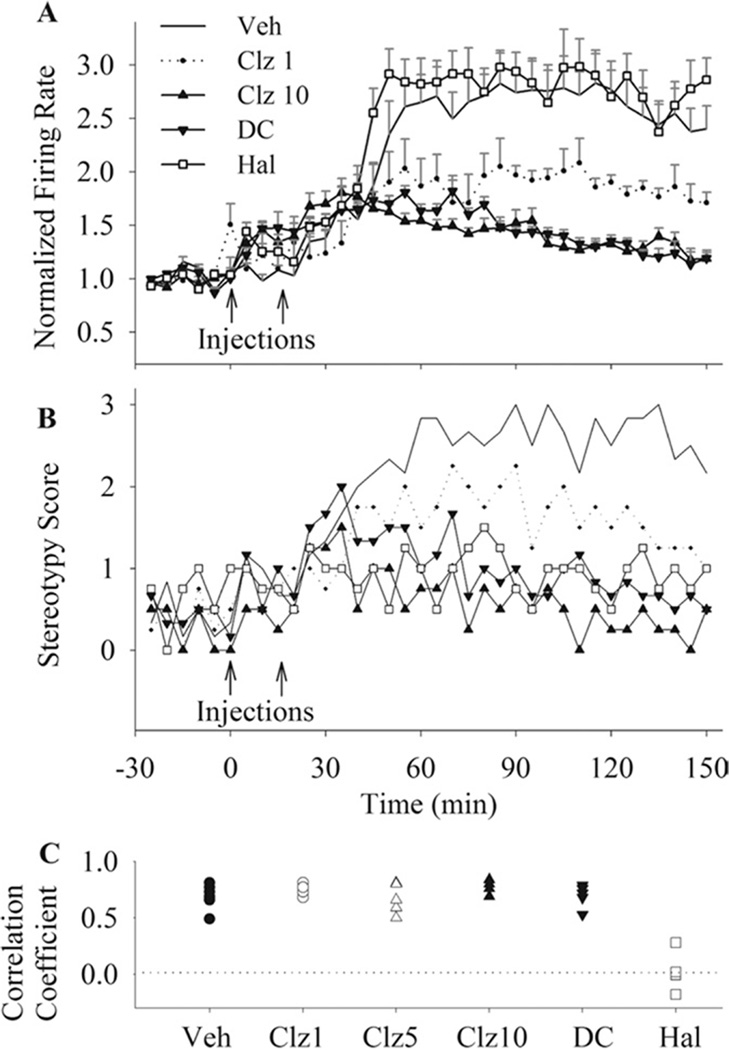
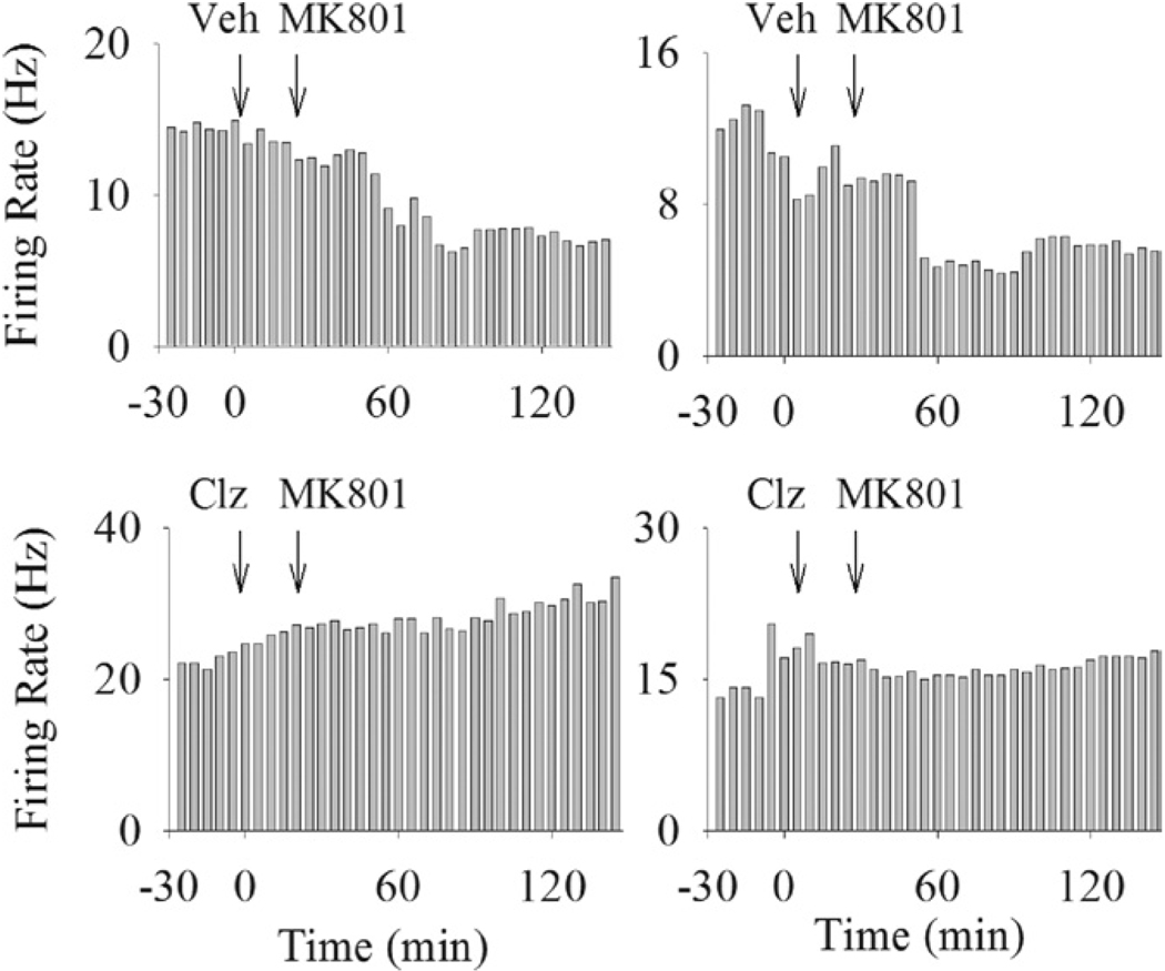
Results: Clozapine and DMClz but not haloperidol had an activity-dependent influence on spontaneous firing rate of PFC cells: they increased the activity of neurons with low baseline firing rates and decreased the activity of neurons with higher firing rates. Clozapine and DMClz but not haloperidol also reversed the effect of MK801 on PFC neuronal firing. This reversal was strongly correlated with blockade of MK801-induced behavioral stereotypy.
Conclusions: These findings indicate that clozapine has the capacity to fine-tune spontaneous and disrupted activity of PFC neurons. This effect might contribute, in part, to the therapeutic efficacy of clozapine in schizophrenia.
Figures
Similar articles
-
Haloperidol and clozapine increase neural activity in the rat prefrontal cortex.Neurosci Lett. 2001 Feb 9;298(3):217-21. doi: 10.1016/s0304-3940(00)01765-1. Neurosci Lett. 2001. PMID: 11165445
-
Antipsychotic drugs reverse the disruption in prefrontal cortex function produced by NMDA receptor blockade with phencyclidine.Proc Natl Acad Sci U S A. 2007 Sep 11;104(37):14843-8. doi: 10.1073/pnas.0704848104. Epub 2007 Sep 4. Proc Natl Acad Sci U S A. 2007. PMID: 17785415 Free PMC article.
-
Effects of clozapine, haloperidol and iloperidone on neurotransmission and synaptic plasticity in prefrontal cortex and their accumulation in brain tissue: an in vitro study.Neuroscience. 2003;117(3):681-95. doi: 10.1016/s0306-4522(02)00769-8. Neuroscience. 2003. PMID: 12617972
-
Activation of noradrenergic locus coeruleus neurons by clozapine and haloperidol: involvement of glutamatergic mechanisms.Int J Neuropsychopharmacol. 2005 Sep;8(3):329-39. doi: 10.1017/S1461145705005080. Epub 2005 Feb 18. Int J Neuropsychopharmacol. 2005. PMID: 15737250
-
Dissociation of haloperidol, clozapine, and olanzapine effects on electrical activity of mesocortical dopamine neurons and dopamine release in the prefrontal cortex.Neuropsychopharmacology. 2000 Jun;22(6):642-9. doi: 10.1016/S0893-133X(00)00087-7. Neuropsychopharmacology. 2000. PMID: 10788763
Cited by
-
Effects of Ketamine on Frontoparietal Interactions in a Rule-Based Antisaccade Task in Macaque Monkeys.J Neurosci. 2024 Dec 11;44(50):e1018232024. doi: 10.1523/JNEUROSCI.1018-23.2024. J Neurosci. 2024. PMID: 39472063 Free PMC article.
-
NeuropsychopharmARCology: Shaping Neuroplasticity through Arc/ Arg3.1 Modulation.Curr Neuropharmacol. 2025;23(6):650-670. doi: 10.2174/011570159X338335240903075655. Curr Neuropharmacol. 2025. PMID: 39473108 Free PMC article. Review.
-
Clozapine Reverses Dysfunction of Glutamatergic Neurons Derived From Clozapine-Responsive Schizophrenia Patients.Front Cell Neurosci. 2022 Feb 23;16:830757. doi: 10.3389/fncel.2022.830757. eCollection 2022. Front Cell Neurosci. 2022. PMID: 35281293 Free PMC article.
-
Antipsychotic drug effects in schizophrenia: a review of longitudinal FMRI investigations and neural interpretations.Curr Med Chem. 2013;20(3):428-37. doi: 10.2174/0929867311320030014. Curr Med Chem. 2013. PMID: 23157635 Free PMC article. Review.
-
A defined network of fast-spiking interneurons in orbitofrontal cortex: responses to behavioral contingencies and ketamine administration.Front Syst Neurosci. 2009 Nov 3;3:13. doi: 10.3389/neuro.06.013.2009. eCollection 2009. Front Syst Neurosci. 2009. PMID: 20057934 Free PMC article.
References
-
- Arango C, Breier A, McMahon R, Carpenter WJ, Buchanan R. The relationship of clozapine and haloperidol treatment response to prefrontal, hippocampal, and caudate brain volumes. Am J Psychiatry. 2003;160:1421–1427. - PubMed
-
- Arvanov V, Liang X, Schwartz J, Grossman S, Wang R. Clozapine and haloperidol modulate N-methyl-D-aspartate- and non-N-methyl-D-aspartate receptor-mediated neurotransmission in rat prefrontal cortical neurons in vitro. J Pharm Exp Thera. 1997;283:226–234. - PubMed
-
- Arvanov V, Wang R. Clozapine, but not haloperidol, prevents the functional hyperactivity of N-methyl-D-aspartate receptors in rat cortical neurons induced by subchronic administration of phencyclidine. J Pharmacol Exp Ther. 1999;289:1000–1006. - PubMed
-
- Baeg EH, Kim YB, Jang J, Kim HT, Mook-Jung I, Jung MW. Fast spiking and regular spiking neural correlates of fear conditioning in the medial prefrontal cortex of rats. Cereb Cortex. 2001;11:441–451. - PubMed
Publication types
MeSH terms
Substances
Grants and funding
LinkOut - more resources
Full Text Sources
Miscellaneous
