. 2006 Dec 10;312(20):4139-49.
doi: 10.1016/j.yexcr.2006.09.012. Epub 2006 Sep 20.

Endonuclease G promotes cell death of non-invasive human breast cancer cells

Affiliations

Endonuclease G promotes cell death of non-invasive human breast cancer cells

Alexei G Basnakian et al. Exp Cell Res. .

Abstract

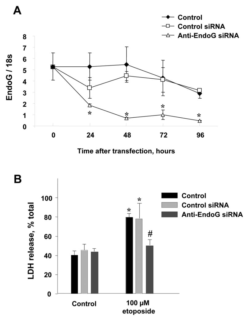
The invasiveness of breast cancer cells was shown to be associated with the suppressed ability to develop apoptosis. The role of cell death DNases/endonucleases has not been previously examined in relation with the invasiveness of breast cancer cells. We have compared the activity of the endonucleases in seven human breast cancer cell lines different in the level of invasiveness and differentiation. The invasiveness of cell lines was confirmed by an in vitro Matrigel-based assay. The total endonuclease activity in the differentiated non-invasive (WDNI) cell lines was higher than that in the poorly differentiated invasive (PDI) cells. The expression of EndoG strongly correlated with the degree of estrogen receptor expression and showed an inverse correlation with vimentin and matrix metalloproteinase-13. The EndoG-positive WDNI cells were more sensitive to etoposide- or camptothecin-induced cell death than EndoG-negative PDI cells. Silencing of EndoG caused inhibited of SK-BR-3 WDNI cell death induced by etoposide. Human ductal carcinomas in situ expressed high levels of EndoG, while invasive medullar and ductal carcinomas had significantly decreased expression of EndoG. This correlated with decreased apoptosis as measured by TUNEL assay. Our findings suggest that the presence of EndoG in non-invasive breast cancer cells determines their sensitivity to apoptosis, which may be taken into consideration for developing the chemotherapeutic strategy for cancer treatment.


Figures

Fig 1
Fig 1
Mn-dependent endonuclease (EndoG) is the second major endonuclease in normal breast epithelium as determined by the inactivation of DNase I. (A) Ca/Mg-dependent endonuclease activity in total extracts from normal breast tissue isolated from DNase I knockout and wild-type mice (n = 4 in each group, *P < 0.01) as measured by PIA. (B) After inactivation of DNase I, a Mn-dependent activity is the most prominent in extracts from DNase I KO mice. Cont, control non-digested pBR322 DNA; Ca, 2 mM CaCl2, pH 7.5; Mg, 2 mM MgCl2, pH 7.5; CM, 2 mM CaCl2 + 2 mM MgCl2,; Mn, 2 mM MnCl2, pH 7.5; CMZ, 2 mM CaCl2 + 2 mM MgCl2 + 2 mM ZnCl2; E5, 2 mM EDTA, no cations, pH 5. Oc-DNA, open circular DNA (relaxed rings with one or more single strand breaks); l-DNA, linear DNA (plasmid linearized by a single double strand break); ccc-DNA, circular covalently closed DNA (intact plasmid without breaks), d-DNA, digested DNA.
Fig 2
Fig 2
Total endonuclease activity and DNase I in breast cancer cells. (A) Endonuclease activity in total protein extracts from human breast cancer cells (top panel) and extracellular activity released into serum-free culture medium after 24 h incubation (bottom panel) as measured using PIA. (B) DNase I zymogram gel electrophoresis of total protein extracts from breast cancer cells (left panel) and tumor xenografts (right panel).
Fig 3
Fig 3
EndoG expression in poorly differentiated invasive breast cancer cells is lower compared to well-differentiated non-invasive cells. (A) Cation requirements of endonuclease in total protein extract from breast cancer cells (SK-BR-3 cells) measured by PIA. Cont, control non-digested pBR322 DNA; Ca, 2 mM CaCl2, pH 7.5; Mg, 2 mM MgCl2, pH 7.5; CM, 2 mM CaCl2 + 2 mM MgCl2,; Mn, 2 mM MnCl2, pH 7.5; CMZ, 2 mM CaCl2 + 2 mM MgCl2 + 2 mM ZnCl2; E5, 2 mM EDTA, no cations, pH 5. The endonuclease that is active in breast cancer cells is not DNase I, but rather a Mn-dependent endonuclease, which is most likely EndoG. (B) EndoG Western blotting of total protein extracted from the cells. (C) Quantification of the EndoG by Western blotting (n = 4 in each group).
Fig 4
Fig 4
Cytotoxicity camptothecin (A) and etoposide (B) to breast cancer cells depends on the presence of EndoG. Well-differentiated non-invasive EndoG-positive cell lines are more sensitive to camptothecin and etoposide. Cells were treated with 2–50 μM camptothecin or 100–300 μM etoposide and cell death was measured by LDH release assay (n = 4 in each group, *p<0.01−0.001).
Fig 5
Fig 5
Silencing of EndoG protects against the cell death of SK-BR-3 breast cancer cells induced by etoposide. (A) Silencing of EndoG was monitored by real-time PCR as described in Methods at 24, 48, 72 and 96 h after transfection (n = 3 in each group, * - p<0.05 in comparison to untreated or control siRNA-treated cells). (B). SK-BR-3 cells were pretreated with specific anti-EndoG and control siRNA for 72h and then exposed to 100 uM etoposide for 24 hours. Cell death was measured by LDH release assay (n = 4 in each group, * - p<0.05 in comparison to untreated control cells; # - p<0.05 in comparison to etoposide-treated cells).
Fig 6
Fig 6
Invasive breast cancer cell lines are EndoG-negative. (A) Comparison of breast cancer markers expression in the used cell lines as measured using cell ELISA. (B) The invasiveness of the cells was determined using the in vitro invasiveness assay, and EndoG was immunostained using anti-EndoG as described in Materials and Methods.
Fig 7
Fig 7
EndoG expression and DNA fragmentation correlate with each other and are decreased in invasive human breast tumors. (A, B) Human ductal carcinomas in situ expressed high levels of EndoG, while invasive medullar and ductal carcinomas had significantly decreased expression of EndoG. Apoptotic DNA fragmentation measured by using TUNEL was present in EndoG positive ductal carcinomas in situ. (C) DNA fragmentation correlated with the EndoG expression.

References

    1. Thompson EW, Paik S, Brunner N, Sommers CL, Zugmaier G, Clarke R, Shima TB, Torri J, Donahue S, Lippman ME. Association of increased basement membrane invasiveness with absence of estrogen receptor and expression of vimentin in human breast cancer cell lines. J Cell Physiol. 1992;150:534–44. - PubMed
    1. Meyer JS, Koehm SL, Hughes JM, Higa E, Wittliff JL, Lagos JA, Manes JL. Bromodeoxyuridine labeling for S-phase measurement in breast carcinoma. Cancer. 1993;71:3531–40. - PubMed
    1. Kim MS, Kang HJ, Moon A. Inhibition of invasion and induction of apoptosis by curcumin in H-ras-transformed MCF10A human breast epithelial cells. Arch Pharm Res. 2001;24:349–54. - PubMed
    1. Fernandez Y, Gu B, Martinez A, Torregrosa A, Sierra A. Inhibition of apoptosis in human breast cancer cells: role in tumor progression to the metastatic state. Int J Cancer. 2002;101:317–26. - PubMed
    1. Mommers EC, van Diest PJ, Leonhart AM, Meijer CJ, Baak JP. Balance of cell proliferation and apoptosis in breast carcinogenesis. Breast Cancer Res Treat. 1999;58:163–9. - PubMed

Publication types