. 2007 Feb 15;109(4):1643-52.
doi: 10.1182/blood-2006-05-023804. Epub 2006 Oct 17.

Prolonged exposure to FLT3 inhibitors leads to resistance via activation of parallel signaling pathways

Affiliations

Prolonged exposure to FLT3 inhibitors leads to resistance via activation of parallel signaling pathways

Obdulio Piloto et al. Blood. .

Abstract

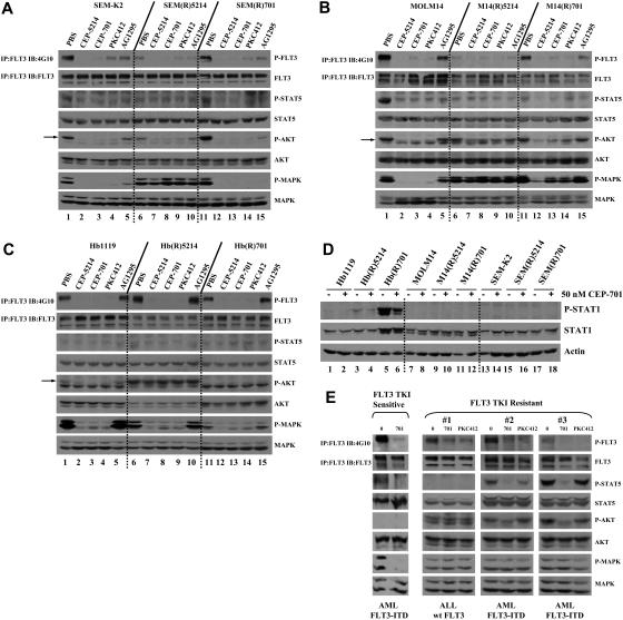
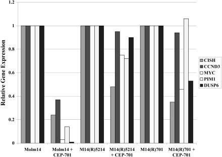
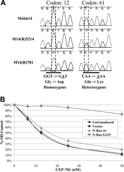
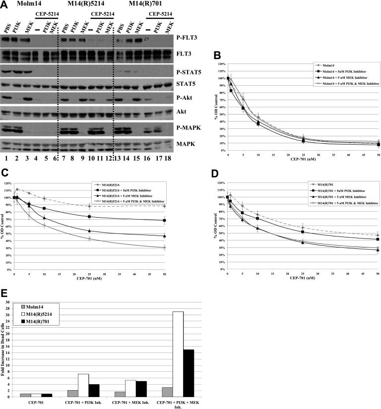
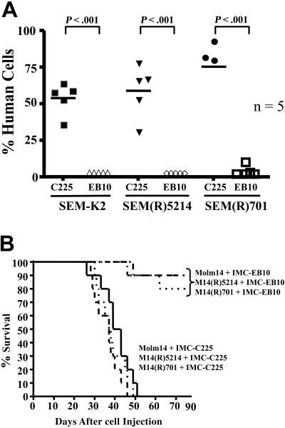
Continuous treatment of malignancies with tyrosine kinase inhibitors (TKIs) may select for resistant clones (ie, imatinib mesylate). To study resistance to TKIs targeting FLT3, a receptor tyrosine kinase that is frequently mutated in acute myelogenous leukemia (AML), we developed resistant human cell lines through prolonged coculture with FLT3 TKIs. FLT3 TKI-resistant cell lines and primary samples still exhibit inhibition of FLT3 phosphorylation on FLT3 TKI treatment. However, FLT3 TKI-resistant cell lines and primary samples often show continued activation of downstream PI3K/Akt and/or Ras/MEK/MAPK signaling pathways as well as continued expression of genes involved in FLT3-mediated cellular transformation. Inhibition of these signaling pathways restores partial sensitivity to FLT3 TKIs. Mutational screening of FLT3 TKI-resistant cell lines revealed activating N-Ras mutations in 2 cell lines that were not present in the parental FLT3 TKI-sensitive cell line. Taken together, these data indicate that FLT3 TKI-resistant cells most frequently become FLT3 independent because of activation of parallel signaling pathways that provide compensatory survival/proliferation signals when FLT3 is inhibited. Anti-FLT3 mAb treatment was still cytotoxic to FLT3 TKI-resistant clones. An approach combining FLT3 TKIs with anti-FLT3 antibodies and/or inhibitors of important pathways downstream of FLT3 may reduce the chances of developing resistance.


Figures

Figure 1
Figure 1
M14(R)5214 and M14(R)701 are resistant to CEP-5214-, CEP-701-, PKC412-, and AG1295-induced apoptosis compared with the parent Molm14 cell line. (A-D) Cells (300 000 cells/mL) were incubated with 0 to 50 nM CEP-5214 or CEP-701, 0 to 100 nM PKC412, or 0 to 20 μM AG1295 for 48 hours at 37°C. Induction of apoptosis was assessed by measuring annexin V binding by flow cytometry. Values indicate percentage of annexin V positive above the untreated samples, which were 5% to 15% annexin V positive. The data are representative of 2 independent experiments.
Figure 2
Figure 2
FLT3 phosphorylation in FLT3 TKI-sensitive and -resistant cell lines and primary samples decreases with FLT3 TKI treatment, although STAT5 and/or Akt and/or MAPK pathways remain activated in most resistant cells. Cell lines expressing (A) wt FLT3 [SEM-K2], (B) FLT3-ITD [MOLM14], or (C) FLT3(D835H) [Hb1119] were treated with PBS, 50 nM CEP-5214, 50 nM CEP-701, 50 nM PKC412, or 1 μM AG1295 for 1 hour at 37°C. Arrow points to P-Akt. (D) Indicated cell lines were treated with PBS or 50 nM CEP-701 for 1 hour at 37°C. (E) Primary samples (20 × 106 cells) were treated with PBS, 50 nM CEP-701, or 50 nM PKC412 for 1 hour at 37°C. Immunoprecipitates and total protein extracts were resolved by 8% SDS–polyacrylamide gel electrophoresis (PAGE) or 10% SDS-PAGE, respectively, and subjected to immunoblot analysis with the indicated phospho-specific antibodies. The same blots were then stripped and reprobed with protein-specific antibodies.
Figure 3
Figure 3
FLT3 TKI-resistant M14(R)5214 and M14(R)701 cell lines maintain higher expression levels of 5 genes regulated by FLT3 signaling and required for FLT3-mediated cellular transformation despite FLT3 inhibition. Molm14, M14(R)5214, and M14(R)701 cell lines were treated with 50 nM CEP-701 for 4 hours prior to isolation of total RNA. The RNA was converted into cDNA and used for Q-PCR analysis. Target gene expression was normalized to GAPDH.
Figure 4
Figure 4
Activating N-Ras mutations were found in the M14(R)5214 and M14(R)701 cell lines and play a role in resistance to FLT3 TKIs. (A) Sequence analysis of cDNA from the Molm14, M14(R)5214, and M14(R)701 cell lines. (B) Molm14 and Molm14 cells (300 000 cells/mL) transduced with the lentiviral vector, wt N-Ras or N-Ras G12V, were treated with CEP-701 for 48 hours at 37°C. Cellular activity/proliferation was determined using the MTT assay in triplicates. Error bars represent standard error of the mean (SEM).
Figure 5
Figure 5
FLT3 TKI-resistant cell lines overexpress a number of RTKs compared with FLT3 TKI-sensitive cell lines. The RNA isolated from FLT3 TKI-sensitive and -resistant cell lines was converted into cDNA and used for Q-PCR analysis in triplicates. Target gene expression was normalized to GAPDH. Error bars indicate SEM.
Figure 6
Figure 6
Resistance to FLT3 TKIs is partially mediated through activation of Akt and/or MAPK signaling pathways. (A) Molm14, M14(R)5214, and M14(R)701 cells (10 × 106 cells) were treated with 5 μM PI3K or MEK inhibitors and/or 50 nM CEP-5214 for 1 hour at 37°C. Immunoprecipitates and total protein extracts were resolved by 8% SDS-PAGE or 10% SDS-PAGE, respectively, and subjected to immunoblot analysis with the indicated phospho-specific antibodies. The same blots were then stripped and reprobed with protein-specific antibodies. (B) Molm14, (C) M14(R)5214, and (D) M14(R)701 cells (300 000 cells/mL) were incubated with 0 to 50 nM CEP-701 and/or 5 μM PI3K or MEK inhibitors for 48 hours at 37°C. Cellular activity/proliferation was determined using the MTT assay in triplicates and normalized to control cells that were not treated with CEP-701. (E) Cells (300 000 cells/mL) were incubated with 25 nM CEP-701 and/or 5 μM PI3K inhibitor and/or 5 μM MEK inhibitor for 48 hours at 37°C. Induction of apoptosis was assessed by measuring annexin V/7-AAD binding by flow cytometry. Values indicate relative fold increase in the percentage of cells that were annexin V/7-AAD positive. Results are representative of 2 independent experiments. Error bars indicate SEM.
Figure 7
Figure 7
IMC-EB10 treatment still reduces leukemic engraftment and prolongs the survival of NOD/SCID mice injected with FLT3 TKI-sensitive or -resistant cell lines. (A) NOD/SCID mice, in groups of 5, were injected with 0.5 × 10⁶ SEM-K2, SEM(R)5214, or SEM(R)701 cells via tail-vein injection. Starting 24 hours after cell injection, mice were injected intraperitoneally a total of 3 times with 400 μg IMC-C225 or IMC-EB10, given every other day. Mice were killed 30 days after cell injection, and bone marrow cells were obtained from femurs. Cells harvested from femurs were stained with hCD19-FITC, mCD45-PE, and hCD45-APC, and human engraftment was determined by flow cytometry. (B) NOD/SCID mice, in groups of 10, were injected with 0.5 × 10⁶ Molm14, M14(R)5214, or M14(R)701 cells via tail-vein injection. Starting 24 hours after cell injection, mice were injected intraperitoneally a total of 3 times with 400 μg IMC-C225 or IMC-EB10, given every other day. Mice were monitored daily for survival.

References

    1. Levis M, Small D. FLT3: ITDoes matter in leukemia. Leukemia. 2003;17:1738–1752. - PubMed
    1. Brown P, Small D. FLT3 inhibitors: a paradigm for the development of targeted therapeutics for paediatric cancer. Eur J Cancer. 2004;40:707–724. - PubMed
    1. Gilliland DG, Griffin JD. The roles of FLT3 in hematopoiesis and leukemia. Blood. 2002;100:1532–1542. - PubMed
    1. Stirewalt DL, Radich JP. The role of FLT3 in haematopoietic malignancies. Nat Rev Cancer. 2003;3:650–665. - PubMed
    1. Nakao M, Yokota S, Iwai T, et al. Internal tandem duplication of the flt3 gene found in acute myeloid leukemia. Leukemia. 1996;10:1911–1918. - PubMed

Publication types