. 2007 Feb 1;109(3):1174-81.
doi: 10.1182/blood-2006-04-015172. Epub 2006 Oct 17.

Massive infection and loss of CD4+ T cells occurs in the intestinal tract of neonatal rhesus macaques in acute SIV infection

Affiliations

Massive infection and loss of CD4+ T cells occurs in the intestinal tract of neonatal rhesus macaques in acute SIV infection

Xiaolei Wang et al. Blood. .

Abstract

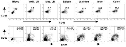
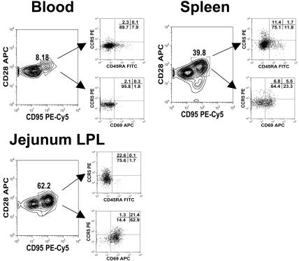
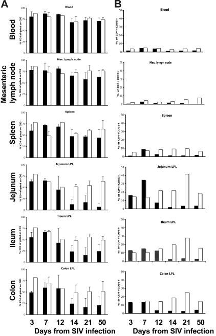
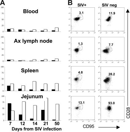
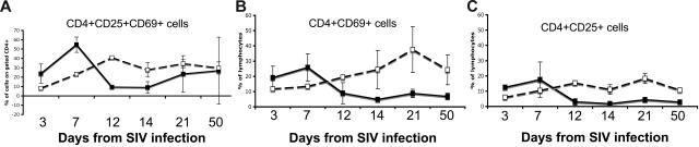
Rapid, profound, and selective depletion of memory CD4+ T cells has now been confirmed to occur in simian immunodeficiency virus (SIV)-infected adult macaques and human immunodeficiency virus (HIV)-infected humans. Within days of infection, marked depletion of memory CD4+ T cells occurs primarily in mucosal tissues, the major reservoir for memory CD4+ T cells in adults. However, HIV infection in neonates often results in higher viral loads and rapid disease progression, despite the paucity of memory CD4+ T cells in the peripheral blood. Here, we examined the immunophenotype of CD4+ T cells in normal and SIV-infected neonatal macaques to determine the distribution of naive and memory T-cell subsets in tissues. We demonstrate that, similar to adults, neonates have abundant memory CD4+ T cells in the intestinal tract and spleen and that these are selectively infected and depleted in primary SIV infection. Within 12 days of SIV infection, activated (CD69+), central memory (CD95+CD28+) CD4+ T cells are marked and persistently depleted in the intestine and other tissues of neonates compared with controls. The results in dicate that "activated" central memory CD4+ T cells are the major target for early SIV infection and CD4+ T cell depletion in neonatal macaques.


Figures

Figure 1
Figure 1
Flow cytometry dot plots demonstrate coexpression of CD95 and CD28 (top) and CD25 and CD69 expression (bottom) on CD4+ T cells from a representative neonate (GB14, 7 days old). Like adults (data not shown), neonates have large numbers of memory (CD95+) and activated (CD25+CD69+) CD4+ T lymphocytes in the intestinal tract (jejunum, ileum, colon) compared with peripheral lymphoid tissues (spleen, lymph nodes, and blood). Central memory cells are defined by CD95 and CD28 coexpression (top, polygonal gates). Note that there are no effector memory cells (CD95+, CD28) evident in any tissues. Plots were generated by gating on CD4+ lymphocytes.
Figure 2
Figure 2
Polychromatic (8 color) flow cytometry demonstrating that central memory (CD95+CD28+) CD4+ T cells from different tissues have different expression patterns of activation markers (CD25, CD69) as well as CCR5 and CD45RA. Plots were gated through lymphocytes and then through CD3+CD4+ T cells from tissues from an uninfected neonate (GA98, 14 days). Note that central memory (CD95+CD28+) CD4+ T cells from the intestine have much higher CD69 and CCR5 expression than those from the spleen or blood.
Figure 3
Figure 3
Bar charts demonstrating percentages of total CD4+ T cells and memory (CD95+) CD4+ T cells in various tissues of SIV-infected and uninfected age-matched normal neonates at various stages of acute infection or age. Note that when examining the changes in total CD4+ T cells (A) there is marked depletion of CD4+ T cells in the intestine (jejunum, ileum, and colon) by 14 days of SIV infection, but minimal changes in CD4+ T cells occur in the blood, lymph nodes, or spleen (A). The selective loss of intestinal CD4+ T cells is a direct reflection of the percentage of memory cells in these tissues before infection (B). Note that intestinal tissues have much higher percentages of memory CD4+ T cells before infection and that these are selectively eliminated in acute SIV infection (B). Data represent percentages of CD4+ cells gated through CD3+ T cells (A) or CD4+CD95+ T cells gated through lymphocytes (B) from groups (standard error bars) or individual animals as described in Table 1. SIV-infected (■) and uninfected (□) age-matched normal neonates.
Figure 4
Figure 4
Changes in central memory CD4+ T cells in neonatal tissues on SIV infection. Bar charts (A) indicate percentages of central memory (CD95+CD28+) CD4+ T cells in SIV-infected (solid bars) to age-matched control neonatal macaques (open bars) over time. Note that a selective loss of central memory CD4+ T cells occurs in the jejunum of SIV-infected neonates by 12 days of infection compared with age-matched controls. Bars represent the percentage of CD95+CD28+ cells after gating through CD4+ T lymphocytes as demonstrated in panel B. (B) Dot plots of CD4+ T cells from tissues of a representative SIV-infected neonate (DP53, left column) to an age-matched uninfected neonate (DP44, right column), demonstrating selective loss of CD95+CD28+ CD4+ T cells in all tissues. Plots were generated by gating through lymphocytes and then CD4+ T cells.
Figure 5
Figure 5
Comparison of CD4+ T cells from the jejunum of neonates. Expression of both CD25 and CD69 (A), CD69 alone (B), or CD25 alone (C) in SIV-infected neonates (solid lines) was compared with age-matched controls (dotted lines). Note that there is a marked and selective loss of activated (CD25+ and/or CD69+) cells in the intestine of all SIV-infected neonates by 12 days of infection.
Figure 6
Figure 6
Immunohistochemistry using monoclonal antibody OPD4 demonstrating memory (CD45RO+) CD4+ T cells in the jejunum of an uninfected neonate (21 days old, DP53) and age-matched SIV-infected neonate (21 days after infection, EC73). Note that numerous memory CD4+ T cells are evident in the uninfected jejunum (A) as compared with the SIV-infected neonate (B). (C) Fluorescent in situ hybridization for SIV combined with immunofluorescence for OPD4 (CD4+ memory cells) in the intestine of 3 SIV-infected neonates examined 12 to 21 days after SIV infection. Essentially all of the SIV-infected cells (red) are CD4+CD45RO+ memory cells as defined by reactivity with monoclonal antibody OPD4 (green) using confocal microscopy. (D) Comparison of absolute numbers of CD4+CD45RO+ cells per square millimeter of jejunum lamina propria between SIV-infected and uninfected (control) neonates. Memory CD4+ T cells are markedly decreased in the intestine of all neonates as early as 12 days after SIV infection. Panels A and B were photographed using a Leica DM LB microscope (Leica Microsystems, Bannockburn, IL) with an N-planar 20×/0.40 NA objective equipped with a Spot 3.2.0 digital camera (Diagnostic Instruments, Sterling Heights, MI). Panel C was taken with a Leica TCS SP2 confocal microscope (Leica Microsystems, Exton, PA). NIH image version 1.62 (NIH, Gaithersburg, MD) and Adobe Photoshop (Adobe Systems, San Jose, CA) were used to assign colors to the images.

References

    1. Veazey RS, DeMaria M, Chalifoux LV, et al. Gastrointestinal tract as a major site of CD4+ T cell depletion and viral replication in SIV infection. Science. 1998;280:427–431. - PubMed
    1. Veazey RS, Mansfield KG, Tham IC, et al. Dynamics of CCR5 expression by CD4(+) T cells in lymphoid tissues during simian immunodeficiency virus infection. J Virol. 2000;74:11001–11007. - PMC - PubMed
    1. Veazey RS, Lackner AA. HIV swiftly guts the immune system. Nat Med. 2005;11:469–470. - PubMed
    1. Mattapallil JJ, Douek DC, Hill B, Nishimura Y, Martin M, Roederer M. Massive infection and loss of memory CD4(+) T cells in multiple tissues during acute SIV infection. Nature. 2005;434:1093–1097. - PubMed
    1. Li Q, Duan L, Estes JD, et al. Peak SIV replication in resting memory CD4(+) T cells depletes gut lamina propria CD4(+) T cells. Nature. 2005;434:1148–1152. - PubMed

Publication types