. 2007 Mar;292(3):F1044-53.
doi: 10.1152/ajprenal.00175.2006. Epub 2006 Oct 17.

Serotonergic drugs and spinal cord transections indicate that different spinal circuits are involved in external urethral sphincter activity in rats

Affiliations

Serotonergic drugs and spinal cord transections indicate that different spinal circuits are involved in external urethral sphincter activity in rats

Hui-Yi Chang et al. Am J Physiol Renal Physiol. 2007 Mar.

Abstract

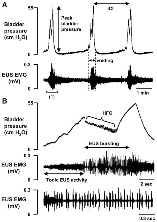
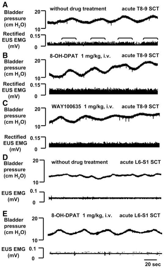
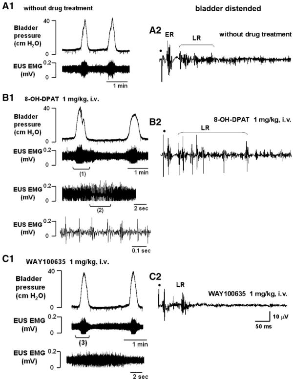
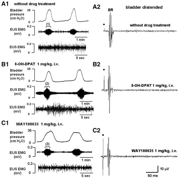
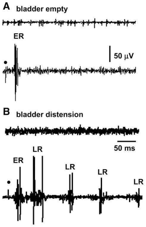
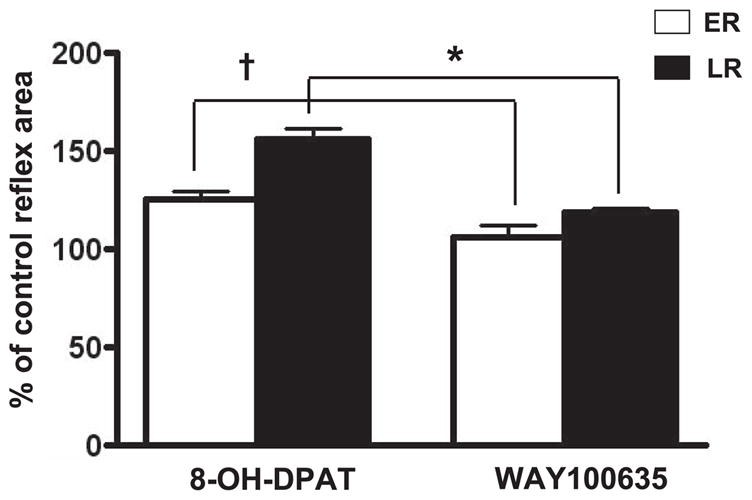
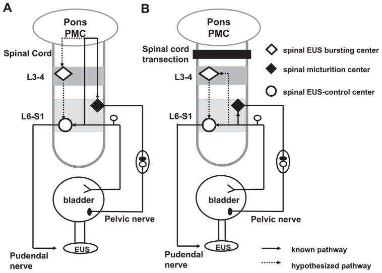
Lower urinary tract function is regulated by spinal and supraspinal reflexes that coordinate the activity of the urinary bladder and external urethral sphincter (EUS). Two types of EUS activity (tonic and bursting) have been identified in rats. This study in urethane-anesthetized female rats used cystometry, EUS electromyography, spinal cord transection (SCT) at different segmental levels, and analysis of the effects of 5-HT(1A) receptor agonist (8-OH-DPAT) and antagonist (WAY100635) drugs to examine the origin of tonic and bursting EUS activity. EUS activity was elicited by bladder distension or electrical stimulation of afferent axons in the pelvic nerve (pelvic-EUS reflex). Tonic activity evoked by bladder distension was detected in spinal cord-intact rats and after acute and chronic T8-9 or L3-4 SCT but was abolished after L6-S1 SCT. Bursting activity was abolished by all types of SCT except chronic T8-9 transection. 8-OH-DPAT enhanced tonic activity, and WAY100635 reversed the effect of 8-OH-DPAT. The pelvic-EUS reflex consisted of an early response (ER) and late response (LR) when the bladder was distended in spinal cord-intact rats. ER remained after acute or chronic T8-9 and L3-4 SCT, but was absent after L6-S1 SCT. LR occurred only in chronic T8-9 SCT rats where it was enhanced or unmasked by 8-OH-DPAT. The results indicate that spinal serotonergic mechanisms facilitate tonic and bursting EUS activity. The circuitry for generating different patterns of EUS activity appears to be located in different segments of the spinal cord: tonic activity at L6-S1 and bursting activity between T8-9 and L3-4.


Figures

Fig. 1
Fig. 1
Bladder pressure (top traces) and external urethral sphincter (EUS) electromyography (EMG) activity (bottom traces) recorded in the rat with intact spinal cord. A: bladder pressure gradually increased during bladder filling at the rate of 0.123 ml/min. A large increase in bladder pressure, which indicates the start of micturition, was accompanied by large-amplitude EUS EMG activity. The bladder pressure consisted of a biphasic response: an initial rise in pressure followed by a decline and then a late secondary rise in pressure. The large-amplitude EUS EMG activity was followed by a prolonged after-discharge that continued for at least half of the intercontraction interval (ICI). B: record at a faster sweep of the time period indicated in A as “(1).” Before the onset of voiding when the bladder was inactive, the EUS exhibited tonic activity. At the beginning of the micturition reflex, tonic EUS activity increased and then converted to bursting activity as bladder pressure declined at the start of voiding. High-frequency oscillations (HFO) occurred in the bladder pressure recording during voiding. Bottom trace shows a fast sweep of the bursting period and conversion of tonic EUS activity to bursting activity at the beginning of voiding.
Fig. 2
Fig. 2
Effects of serotonergic drugs on small-amplitude bladder contractions (top traces) and rectified tonic EUS EMG activity (bottom traces) in 2 rats after T8–9 (AC) and L6–S1 (D and E) acute spinal cord transection (SCT). A: a small increase in tonic EUS activity occurred during bladder contractions (brackets), but EUS bursting activity did not occur. B: after 8-OH-DPAT, the amplitude of tonic EUS activity increased but bladder contractions did not significantly change. C: WAY100635 reduced the facilitatory effect of 8-OH-DPAT on tonic EUS activity. Neither 8-OH-DPAT nor WAY100635 affected small-amplitude bladder contractions. The interval between administration of 8-OH-DPAT and WAY100635 was 30 min. All records (AC) were obtained in the same animal. D and E: acute L6–S1 SCT eliminated EUS EMG activity and the effects of 8-OH-DPAT.
Fig. 3
Fig. 3
Effects of serotonergic drugs on the bladder (top left traces), EUS EMG activity (bottom left traces), and pelvic-EUS reflex (right traces) in a rat with T8–9 chronic SCT (4 wk). A1: before drug treatment, the bladder exhibited rhythmic, large-amplitude micturition contractions during bladder filling. During the bladder contractions, the low-amplitude tonic EUS activity was enhanced but EUS bursting did not occur. A2: early response (ER) and a small-amplitude late response (LR) in the pelvic-EUS reflex was present when the bladder was distended. B1: 8-OH-DPAT increased the tonic EUS activity and unmasked EUS bursting during voiding. Trace 3 shows a fast sweep of EUS EMG activity during period (1) that included the bursting. Trace 4 shows a faster sweep of the EUS bursting during period (2) in trace 3. B2: after 8-OH-DPAT, the LR was markedly increased. C1: WAY100635 reversed the effect of 8-OH-DPAT. The large-amplitude tonic activity was reduced and the EUS bursting during voiding was eliminated. Trace 3 is a fast sweep of the period (3) showing that EUS bursting was eliminated by WAY100635, leaving only tonic EUS activity when the bladder pressure increased during a micturition reflex. C2: after WAY100635 the burst firing of the LR was suppressed. Dot, electrical stimulation (4 V, 1.0 Hz, pulse width 0.05 ms). All traces (A2C2) represent a recording of a single reflex response.
Fig. 4
Fig. 4
Effects of serotonergic drugs on the bladder (top left traces), EUS EMG activity (bottom left traces), and pelvic-EUS reflex (right traces) in a rat with chronic L3–4 SCT during bladder filling and repeated voiding. A1, B1, C1: in each record, EUS EMG activity is shown at a slow time base (middle trace) and an expanded time base (bottom trace) of the sequence of recording indicated by the brackets (1, 2, 3) in the middle traces. A1: before drug treatment, the bladder exhibited rhythmic micturition contractions during bladder filling. At the peak of the contractions, tonic EUS activity was markedly enhanced. EUS bursting activity did not occur. B1: 8-OH-DPAT enhanced tonic EUS activity but did not unmask EUS bursting. C1: WAY100635 reversed the effect of 8-OH-DPAT on tonic EUS activity. A2C2: the ER remained, but the LR was absent in the chronic L3–4 SCT rat. The ER was not affected by drugs. Dot, electrical stimulation (6 V, 1.0 Hz, pulse width 0.05 ms).
Fig. 5
Fig. 5
Pelvic-EUS reflex was enhanced by bladder distension in a rat with an intact spinal cord. A: top trace shows the EUS-EMG activity without electrical stimulation. Bottom trace shows the pelvic-EUS reflex elicited by a single shock to the pelvic nerve (dot) when the bladder was empty. Reflex responses consisted of a large ER. B: top trace shows the EUS-EMG activity without electrical stimulation when the bladder was distended by 0.2 ml of saline. Bottom trace shows that the LR of pelvic-EUS reflex was unmasked but the ER was not changed by bladder distension. All traces were obtained in the same animal and represent a recording of a single reflex response. Dot, 4 V, 0.1 Hz, pulse width 0.05 ms.
Fig. 6
Fig. 6
Area of the pelvic-EUS ER and LR reflexes in chronic T8–9 SCT rats when the bladder was distended. The ER and LR were significantly enhanced 26 (P < 0.05) and 56% (P < 0.05), respectively, by 8-OH-DPAT (n = 5). WAY100635 (n = 5) significantly decreased ER and LR by 18 and 38%, respectively, after 8-OH-DPAT. *, †: P < 0.05, significantly decreased compared with records after 8-OH-DPAT.
Fig. 7
Fig. 7
Diagram showing putative reflex pathways mediating reflex micturition and tonic and bursting EUS activity in spinal cord-intact (A) and chronic SCT T8–9 rats (B). A: spinobulbospinal micturition reflex pathway is shown by the solid line passing through the pontine micturition center (PMC) in the rostral brain stem. The hypothesized pathway mediating EUS bursting is shown by the dotted line also passing through the PMC. In spinal cord-intact rats, when the bladder is distended, afferent input from bladder mechanoreceptors passes via the pelvic nerve to the L6–S1 spinal cord to the spinal EUS-control center to generate tonic EUS activity and the ER. Input from the L6–S1 spinal cord passes to the PMC, which then projects to the lumbosacral micturition center to generate reflex bladder contractions and to L3–4 bursting center to generate EUS bursting. The spinal EUS bursting center provides an excitatory input to the spinal EUS-control center to initiate an excitatory outflow to the EUS. The spinal EUS-control center in the L6–S1 spinal cord consists of interneuronal and motoneuronal circuitry that regulates EUS activity. B: after SCT, descending input from the PMC to spinal centers is interrupted. This initially eliminates the micturition reflex, the LR, and EUS bursting. The ER and tonic EUS activity mediated by a spinal reflex pathway are preserved. However, in chronic SCT rats it is hypothesized that reorganization of synaptic connections in the spinal cord leads to the reemergence of the micturition reflex as well as the LR and EUS bursting. This reorganization depends on the formation of new pathways between pelvic primary afferent nerves and the L3–4 spinal EUS bursting center (dotted line) and spinal micturition center (solid line) or upregulation of pathways that exist in the spinal intact animals.

References

    1. Chang HY, Cheng CL, Chen JJ, de Groat WC. Abstract Viewer (Online). Program No. 541.13. Society for Neuroscience; 2004. Role of glutamatergic and serotonergic mechanisms in urethral sphincter reflexes in urethane-anesthetized rats. http://www.sfn.org.
    1. Chang HY, Cheng CL, Chen JJ, Negoita FA, de Groat WC. Abstract Viewer (Online). Program No. 48.17. Society for Neuroscience; 2005. Influence of serotonergic mechanisms in the spinal cord on external urethral sphincter function during voiding. http://www.sfn.org.
    1. Chang HY, Cheng CL, Peng CW, Chen JJ, de Groat WC. Reflexes evoked by electrical stimulation of afferent axons in the pudendal nerve under empty and distended bladder conditions in urethane-anesthetized rats. Neurosci Meth. 2006;150:80–89. - PMC - PubMed
    1. Cheng CL, de Groat WC. The role of capsaicin-sensitive afferent fibers in the lower urinary tract dysfunction induced by chronic spinal cord injury in rats. Exp Neurol. 2004;187:445–454. - PubMed
    1. Cheng CL, Ma CP, de Groat WC. Effect of capsaicin on micturition and associated reflexes in chronic spinal rats. Brain Res. 1995;678:40–48. - PubMed

Publication types

MeSH terms

Substances