. 2006 Oct 23;95(8):1020-7.
doi: 10.1038/sj.bjc.6603363. Epub 2006 Oct 3.

The oncolytic effect in vivo of reovirus on tumour cells that have survived reovirus cell killing in vitro

Affiliations

The oncolytic effect in vivo of reovirus on tumour cells that have survived reovirus cell killing in vitro

T Alain et al. Br J Cancer. .

Abstract

The use of oncolytic viruses has received considerable attention in recent years and many viruses have proved to be effective against a variety of cancer models and a few are currently being used in clinical trials. However, the possible emergence and outcome of virus-resistant tumour cells has not been addressed. We previously reported the effective use of reovirus against lymphoid malignancies, including the Burkitt's lymphoma cell line Raji. Here we isolated in vitro persistently infected (PI) Raji cells, and cells 'cured' of persistent reovirus infection ('cured' cells). Both PI and cured Raji cells resisted reovirus infection and cell killing in vitro. In vivo, the PI cells were non-tumorigenic in SCID mice, but cured cells regained the parental cells' ability to form tumours. Tumour xenografts from the cured cells, however, were highly susceptible to reovirus oncolysis in vivo. This susceptibility was due to the proteolytic environment within tumours that facilitates reovirus infection and cell killing. Our results show that persistent infection by reovirus impedes tumour development and that although PI cells cleared of reovirus are tumorigenic, they are killed upon rechallenge with reovirus. Both the PI and cured states are therefore not likely to be significant barriers to reovirus oncolytic therapy.


Figures
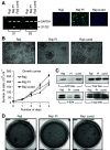
Figure 1
Figure 1
Growth of the Raji parental, PI and cured cells. (A) The left panel shows polymerase chain reaction of reovirus S1 and S2 mRNA transcript in Raji parental, PI and cured cells. Equal amounts of RNA from each sample were subjected to RT–PCR, followed by selective amplification of reovirus S1 or S2 cDNA and GAPDH. The right panel shows immunofluorescence of reovirus proteins expressed only in PI cells. Cells grown under normal conditions were fixed, processed and reacted with rabbit anti-reovirus type 3 antibody, followed by FITC-conjugated goat anti-rabbit IgG and mounted with DAPI-stained mounting medium. The magnification for all panels was × 400. (B) Photomicrographs of the Raji parental, PI and cured cells in culture and growth curves assessed by staining the cells with 0.25% Trypan Blue. Viable (unstained) cells from three independent wells were counted using a haemocytometer. (C) Ras and ERK activity in Raji, PI and cured cells. (D) Growth of Raji parental, PI and cured cells in soft agar. A total of 1 × 10⁵ cells were mixed (1 : 1) in 2 × RPMI containing 10% FBS and 1.2% low-melting temperature agarose (SeaPlaque) and allowed to grow for 4 weeks. Colonies were then fixed, stained with Coomassie brilliant blue and photomicrographed.
Figure 2
Figure 2
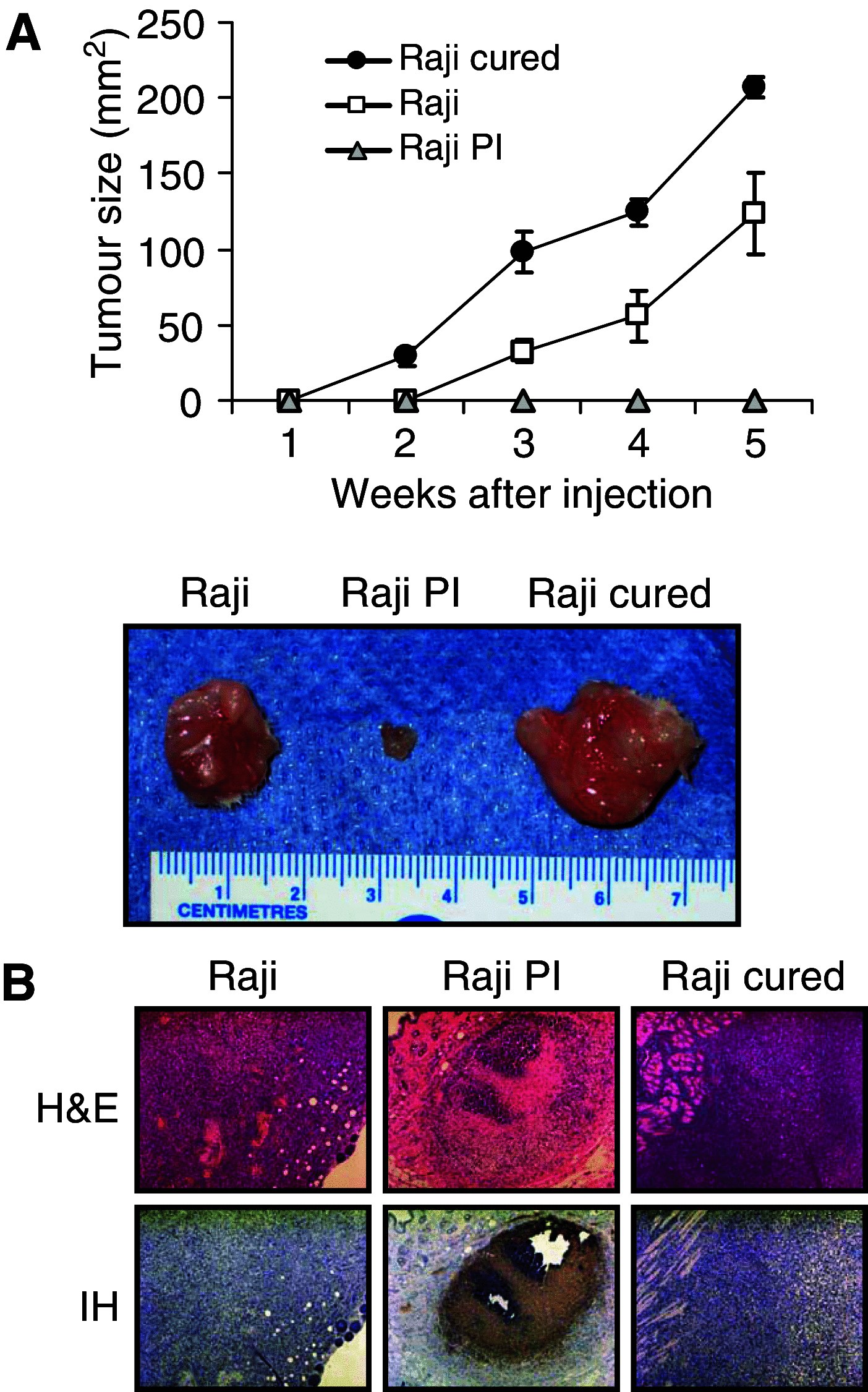
Tumorigenicity of Raji parental, PI and cured cells in SCID mice. (A) SCID mice were subcutaneously implanted with 1 × 10⁷ cells of Raji parental, PI or cured cells. Tumour growth was followed for a period of 5 weeks and two-dimensional measurements were taken weekly with a caliper. Resected tumours were photographed. (B) Haematoxylin and eosin staining and IH of reovirus antigens in tumour tissue taken from Raji parental, PI and cured tumours. Haematoxylin- and eosin-stained section (original magnification × 200) show large tumours 30 days postinjection in the Raji and cured group. Tumours in the PI group were not apparent on palpation and only small foci of tumour were found on histological examination. Immunohistochemical of tumour sections (original magnification × 200) shows that only PI cells stain positively for reovirus proteins (brown), although most of the tumour is necrotic.
Figure 3
Figure 3
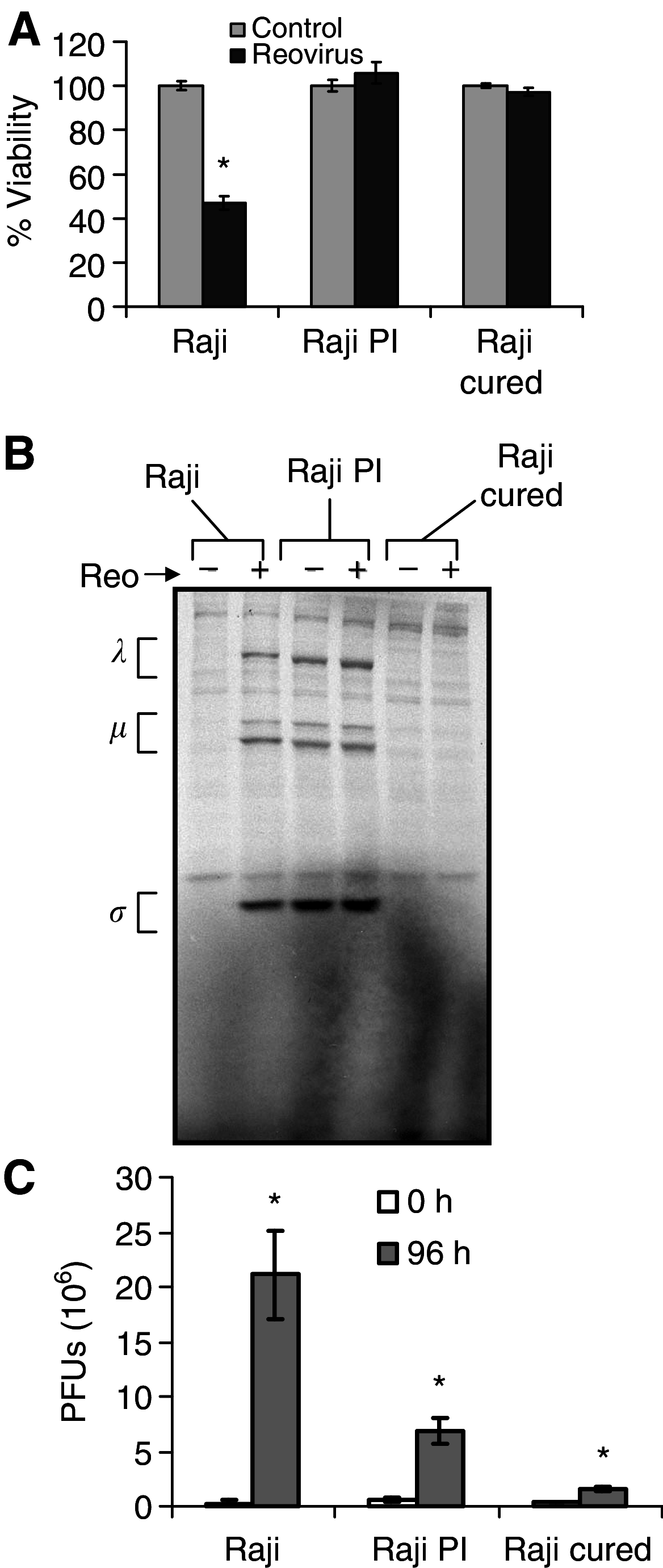
Effect of reovirus on Raji parental and cured cells in vitro. (A) Cell viability. Raji parental, PI and cured cells were exposed to reovirus at an MOI of 20 PFU cell−1, and viability at 96 h postinfection was assessed using WST-1 assay from three independent wells. Compared to controls, P-values for reovirus-treated groups are P<0.001, P=0.93 and P=0.222 for Raji, Raji PI and Raji cured, respectively. (B) Viral protein synthesis. Infected cells were metabolically labelled with [35S]methionine for 6 h at 42 h postinfection. Cell lysates were prepared, and reovirus proteins were immunoprecipitated with a polyclonal anti-reovirus antibody and analysed by SDS–PAGE. The three size classes of reovirus proteins (λ, μ and σ) are indicated on the left. (C) Virus progeny. At 0 and 96 h postinfection, cells were harvested and freeze–thawed three times, and the virus titre in the lysate was determined by plaque assay on L929 cells. Compared to input virus, P-values for progeny virus at 96 h postinfection are P<0.001, P=0.0015 and P<0.001. Error bars indicate s.e.m. from three separate wells and *demonstrates statistical significance.
Figure 4
Figure 4
Effect of reovirus on parental and cured Raji tumours in vivo. (A) Intratumoral reovirus therapy of lymphoid tumours in SCID mice. SCID mice were subcutaneously implanted with 1 × 10⁷ cells of either Raji parental or cured cells. Following palpable tumour establishment, the tumours received (on day 0) a single intratumoral injection of 1 × 10⁷ PFUs of live reovirus (n=8) or saline control (n=7). Tumour growth was followed for a period of 25 days and measured two-dimensionally with a caliper. *Differences are statistically significant with P<0.001 for Raji+reovirus and P=0.002 for Raji cured+reovirus compared to respective controls. (B) Haematoxylin and eosin staining and IH of reovirus antigens in Raji parental and cured tumours after intratumoral reovirus treatment. Haematoxylin- and eosin-stained section shows necrosis of tumour cells 24 days after live reovirus treatment. IH-stained section (original magnification × 100 (left) and × 200 (right)) of remaining tumour cells stains positively for reovirus proteins (brown). Histological examination of saline-treated (control) Raji and cured tumours showed very large actively proliferating tumours (data not shown).
Figure 5
Figure 5
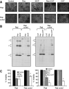
Infection of Raji cured cells by ISVP, or by reovirions in the presence of chymotrypsin. (A) Cytopathic effects. Raji parental or cured cells were exposed to ISVP, reovirus or reovirus in the presence of chymotrypsin (CHT) at an MOI of 20 PFU cell−1, and photomicrographs were taken at 72 h postinfection. Original magnification × 400. (B) Reovirus protein synthesis in infected Raji parental and cured cells. Cells were infected with ISVP, reovirus or reovirus in the presence of CHT, and pulse-labelled with [35S]methionine for 6 h at 18 h postinfection. The cells were then harvested and lysed, and reovirus proteins were immunoprecipitated from an aliquot of the lysate using a rabbit polyclonal anti-reovirus antibody, followed by SDS–PAGE. The three size classes of reovirus proteins (λ, μ and σ) are indicated on the left. (C) Viability of Raji parental and cured cells infected with ISVP, reovirus or reovirus in the presence of CHT. WST-1 assay was carried out on cells from three independent wells at 96 h postinfection. P-values compared with respective uninfected controls are: Left panel, P=0.012 (Raji+Reo), P=0.017 (Raji+ISVP), P=0.61 (Raji cured+Reo) and P=0.026 (Raji cured+ISVP). Right panel, P=0.019 (Raji+Reo), P<0.001 (Raji+Reo+CHT), P=0.98 (Raji cured+Reo) and P=0.0013 (Raji cured+Reo+CHT). *Indicates statistical significance.
Figure 6
Figure 6
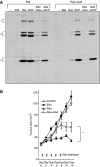
Infection of Raji and Raji cured cells in the presence of E64. (A) Reovirus protein synthesis in infected Raji parental and cured cells treated with E64. Cells were infected with ISVP or reovirus in the presence or absence of E64, and pulse-labelled with [35S]methionine for 6 h at 18 h postinfection. The cells were then harvested and lysed, and reovirus proteins were immunoprecipitated from an aliquot of the lysate using a rabbit polyclonal anti-reovirus antibody, followed by SDS–PAGE. The three size classes of reovirus proteins (λ, μ and σ) are indicated on the left. (B) Intratumoral reovirus therapy of lymphoid tumours in SCID mice exposed to E64 injected i.p. SCID mice were subcutaneously implanted with 1 × 10⁷ cells of Raji parental cells. Following palpable tumour establishment, the tumours received (on day 0) either 1 mg per mouse of E64 in PBS alone (n=5) or 1 h before a single intratumoral injection of 1 × 10⁷ PFUs of live reovirus (n=5) and E64 was re-injected subsequently every second day until day 8. E64 treatments were compared to PBS control and reovirus alone group (n=5). Tumour growth was followed for a period of 12 days and measured two-dimensionally with a caliper. *Wilcoxon's signed-rank test (P=0.0379) comparing tumour sizes of reovirus+E64-treated group to reovirus-alone group.

Similar articles

Cited by

References

    1. Ahmed R, Fields BN (1982) Role of the S4 gene in the establishment of persistent reovirus infection in L cells. Cell 28: 605–612 - PubMed
    1. Ahmed R, Canning WM, Kauffman RS, Sharpe AH, Hallum JV, Fields BN (1981) Role of the host cell in persistent viral infection: coevolution of L cells and reovirus during persistent infection. Cell 25: 325–332 - PubMed
    1. Alain T, Hirasawa K, Pon KJ, Nishikawa SG, Urbanski SJ, Auer Y, Luider J, Martin A, Johnston RN, Janowska-Wieczorek A, Lee PW, Kossakowska AE (2002) Reovirus therapy of lymphoid malignancies. Blood 100: 4146–4153 - PubMed
    1. Amerongen HM, Wilson GA, Fields BN, Neutra MR (1994) Proteolytic processing of reovirus is required for adherence to intestinal M cells. J Virol 68: 8428–8432 - PMC - PubMed
    1. Baer GS, Dermody TS (1997) Mutations in reovirus outer-capsid protein sigma3 selected during persistent infections of L cells confer resistance to protease inhibitor E64. J Virol 71: 4921–4928 - PMC - PubMed

Publication types

MeSH terms