. 2004 May;1(2):157-63.
doi: 10.1017/s1740925x04000249.

Overexpression of c-MYC promotes an undifferentiated phenotype in cultured astrocytes and allows elevated Ras and Akt signaling to induce gliomas from GFAP-expressing cells in mice

Affiliations

Overexpression of c-MYC promotes an undifferentiated phenotype in cultured astrocytes and allows elevated Ras and Akt signaling to induce gliomas from GFAP-expressing cells in mice

Andrew B Lassman et al. Neuron Glia Biol. 2004 May.

Abstract

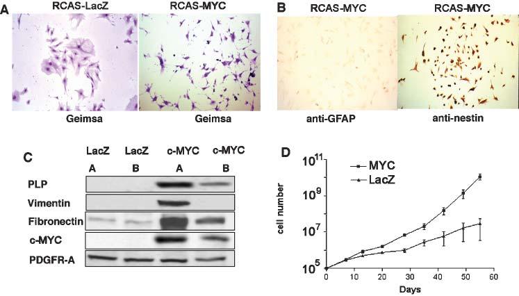
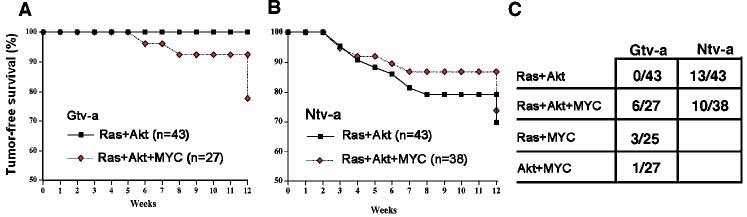
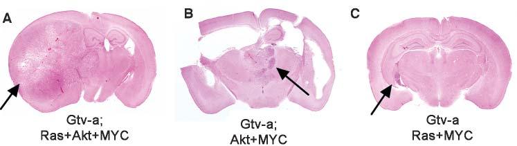
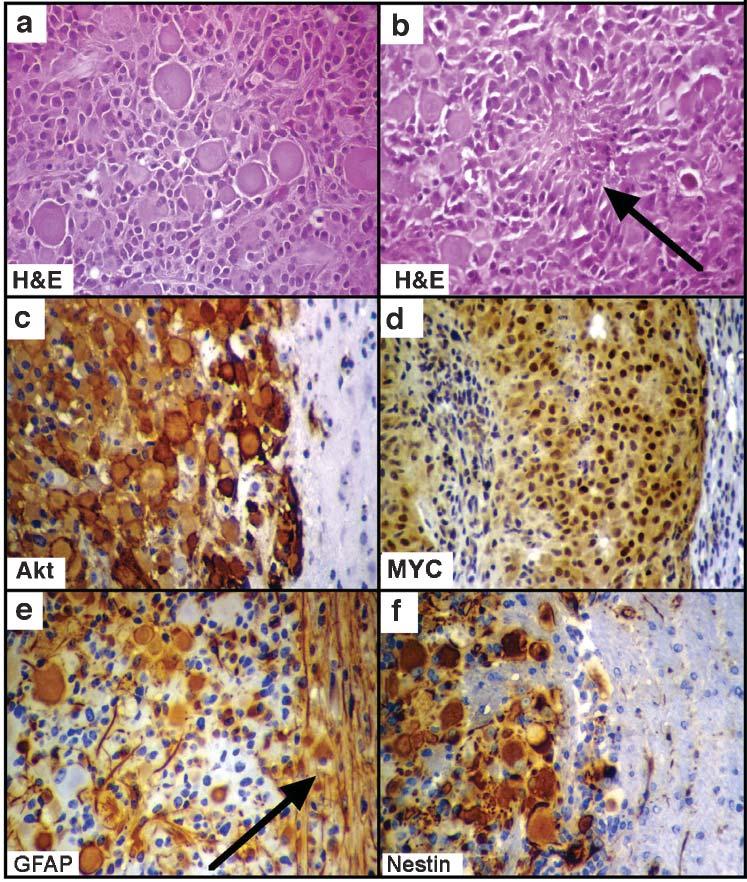
The c-MYC protooncogene is overexpressed in the most malignant primary brain tumor, glioblastoma multiforme (GBM), and has been correlated with the undifferentiated character of several cell types. However, the role of Myc activity in the generation of GBMs is not known. In this report, we show that gene transfer of c-MYC to GFAP-expressing astrocytes in vitro promotes the outgrowth of GFAP-negative, nestin-expressing cells with progenitor-like morphology, growth characteristics and gene-expression pattern. In addition, gene transfer of c-MYC to GFAP-expressing astrocytes in vivo induces GBMs when co-expressed with activated Ras and Akt. Without c-MYC, Ras+Akt induces GBMs from nestin-expressing CNS progenitors but is insufficient in GFAP-expressing differentiated astrocytes. The ability of Myc activity to enhance the oncogenic effects of Ras+Akt appears to be limited to GFAP-expressing astrocytes because nestin-expressing progenitors show no increase in GBM formation with the addition of MYC to Ras+Akt. These studies indicate that one role of MYC activity in the formation of gliomas might be to either promote or reinforce an undifferentiated phenotype required for glioma cells to respond to the oncogenic effects of elevated Ras and Akt activity.


Figures

Fig. 1
Fig. 1
Cell-culture characteristics of astrocytes infected with RCAS-MYC. (A) Morphologic differences between Gtv-a astrocytes infected with RCAS-MYC and RCAS-LacZ are indicated. (B) Immunostaining for either GFAP or nestin in cultured astrocytes from Gtv-a mice infected with RCAS-MYC. (C) Western blot analysis of two, independent, RCAS-MYC-infected Gtv-a astrocyte cultures demonstrating expression of proteins that are not typically expressed in astrocytes but are characteristic of glial progenitors, including PLP, vimentin and fibronectin. (D) Growth rates of RCAS-MYC-infected and RCAS-LacZ (control)-infected Gtv-a transgenic astrocytes in culture. A and B at 100× magnification.
Fig. 2
Fig. 2
Tumor-free survival and tumor incidence in Gtv-a and Ntv-a-transgenic mice infected with RCAS vectors carrying activated Ras, Akt and human c-MYC. Two cohorts each of Gtv-a (A) and Ntv-a (B) transgenic mice were infected with either RCAS-Ras + RCAS-Akt or RCAS-Ras + RCAS-Akt + RCAS-MYC. Mice were sacrificed either at 12 weeks of age or when they developed signs of intracranial pathology. (C) The incidence of tumors in Ntv-a and Gtv-a mice infected with RCAS vectors. The denominator indicates the number of mice infected at birth and the numerator indicates the number of mice that developed tumors by 12 weeks of age.
Fig. 3
Fig. 3
Characteristics of GBMs induced from astrocytes by infection of mice with RCAS-MYC, RCAS-Ras and RCAS-Akt. Whole-mounts illustrating (A) the largest GBM arising in Gtv-a mice after infection with a combination of Ras+Akt+MYC, (B) the only GBM that arose in a Gtv-a mouse after infection with Akt+MYC (with associated hydrocephalus), and (C) the largest GBM arising in Gtv-a mice after infection with Ras+MYC.
Fig. 4
Fig. 4
Microscopic characteristics of gliomas arising from astrocytes following infection of Gtv-a mice with RCAS vectors encoding Ras, Akt and MYC. H&E staining illustrates (A) the mixed cell type and (B) pseudopalisading necrosis (arrow). (C–F) Immunohistochemical staining with antibodies to the HA-tag on the virally transduced Akt (to identify the tumor) (C), human c-MYC (D), GFAP (E) and nestin (F). All photographs of immunohistochemical stains include both tumor and adjacent brain tissue for comparison. The progenitor marker nestin is expressed in the tumor arising from GFAP-expressing cells, and GFAP is expressed in both tumor and adjacent reactive brain tissue (arrow in E). All at 400× magnification.

References

    1. Aoki M, Batista O, Bellacosa A, Tsichlis P, Vogt PK. The Akt kinase: Molecular determinants of oncogenicity. Proceedings of the National Academy of Sciences of the United States of America. 1998;95:14950–14955. - PMC - PubMed
    1. Bigner DD, McLendon RE, Bruner JM, Russell DS, Rubinstein LJ. Russell and Rubinstein's Pathology of Tumors of the Nervous System. Hodder Arnold; 1998.
    1. Chin L, Schreiber-Agus N, Pellicer I, Chen K, Lee HW, Dudast M, Cordon-Cardo C, DePinho RA. Contrasting roles for Myc and Mad proteins in cellular growth and differentiation. Proceedings of the National Academy of Sciences of the United States of America. 1995;92:8488–8492. - PMC - PubMed
    1. Dai C, Celestino JC, Okada Y, Louis DN, Fuller GN, Holland EC. PDGF autocrine stimulation dedifferentiates cultured astrocytes and induces oligodendrogliomas and oligoastrocytomas from neural progenitors and astrocytes in vivo. Genes and Development. 2001;15:1913–1925. - PMC - PubMed
    1. Engelhard HH, 3rd, Butler A.B.t., Bauer KD. Quantification of the c-myc oncoprotein in human glioblastoma cells and tumor tissue. Journal of Neurosurgery. 1989;71:224–232. - PubMed