

Remote non-invasive stereoscopic imaging of blood vessels: first in-vivo results of a new multispectral contrast enhancement technology
- PMID: 17048103
- PMCID: PMC1705509
- DOI: 10.1007/s10439-006-9198-1
Remote non-invasive stereoscopic imaging of blood vessels: first in-vivo results of a new multispectral contrast enhancement technology
Abstract
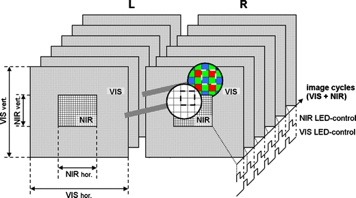
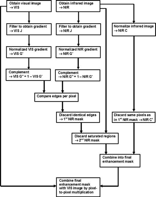
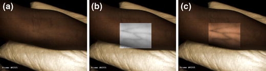
We describe a contactless optical technique selectively enhancing superficial blood vessels below variously pigmented intact human skin by combining images in different spectral bands. Two CMOS-cameras, with apochromatic lenses and dual-band LED-arrays, simultaneously streamed Left (L) and Right (R) image data to a dual-processor PC. Both cameras captured color images within the visible range (VIS, 400-780 nm) and grey-scale images within the near infrared range (NIR, 910-920 nm) by sequentially switching between LED-array emission bands. Image-size-settings of 1280 x 1024 for VIS & 640 x 512 for NIR produced 12 cycles/s (1 cycle = 1 VIS L&R-pair + 1 NIR L&R-pair). Decreasing image-size-settings (640 x 512 for VIS and 320 x 256 for NIR) increased camera-speed to 25 cycles/s. Contrasts from below the tissue surface were algorithmically distinguished from surface shadows, reflections, etc. Thus blood vessels were selectively enhanced and back-projected into the stereoscopic VIS-color-image using either a 3D-display or conventional shutter glasses. As a first usability reconnaissance we applied this custom-built mobile stereoscopic camera for several clinical settings:* blood withdrawal;* vein inspection in dark skin;* vein detection through iodide;* varicose vein and nevi pigmentosum inspection. Our technique improves blood vessel visualization compared to the naked eye, and supports depth perception.
Figures


Similar articles
-
Contrast enhancement of coronary arteries in cardiac surgery: a new multispectral stereoscopic camera technique.EuroIntervention. 2006 Nov;2(3):389-94. EuroIntervention. 2006. PMID: 19755318
-
Scanning multispectral IR reflectography SMIRR: an advanced tool for art diagnostics.Acc Chem Res. 2010 Jun 15;43(6):847-56. doi: 10.1021/ar900268t. Acc Chem Res. 2010. PMID: 20230039
-
Introducing 3-dimensional stereoscopic imaging to the study of musculoskeletal anatomy.Arthroscopy. 2011 Apr;27(4):593-6. doi: 10.1016/j.arthro.2011.01.004. Arthroscopy. 2011. PMID: 21444013 Review.
-
Stereoscopic navigation-controlled display of preoperative MRI and intraoperative 3D ultrasound in planning and guidance of neurosurgery: new technology for minimally invasive image-guided surgery approaches.Minim Invasive Neurosurg. 2003 Jun;46(3):129-37. doi: 10.1055/s-2003-40736. Minim Invasive Neurosurg. 2003. PMID: 12872188
-
PET/CT image navigation and communication.J Nucl Med. 2004 Jan;45 Suppl 1:46S-55S. J Nucl Med. 2004. PMID: 14736835 Review.
Cited by
-
Photoplethysmographic Imaging of Hemodynamics and Two-Dimensional Oximetry.Opt Spectrosc. 2022;130(7):452-469. doi: 10.1134/S0030400X22080057. Epub 2022 Nov 29. Opt Spectrosc. 2022. PMID: 36466081 Free PMC article.
-
Rapid prototyping of biomimetic vascular phantoms for hyperspectral reflectance imaging.J Biomed Opt. 2015;20(12):121312. doi: 10.1117/1.JBO.20.12.121312. J Biomed Opt. 2015. PMID: 26662064 Free PMC article.
-
Remote plethysmographic imaging using ambient light.Opt Express. 2008 Dec 22;16(26):21434-45. doi: 10.1364/oe.16.021434. Opt Express. 2008. PMID: 19104573 Free PMC article.
-
Optical imaging for breast cancer prescreening.Breast Cancer (Dove Med Press). 2015 Jul 20;7:193-209. doi: 10.2147/BCTT.S51702. eCollection 2015. Breast Cancer (Dove Med Press). 2015. PMID: 26229503 Free PMC article. Review.
-
Context-free hyperspectral image enhancement for wide-field optical biomarker visualization.Biomed Opt Express. 2019 Dec 9;11(1):133-148. doi: 10.1364/BOE.11.000133. eCollection 2020 Jan 1. Biomed Opt Express. 2019. PMID: 32010505 Free PMC article.
References
-
- {'text': '', 'ref_index': 1, 'ids': [{'type': 'DOI', 'value': '10.1016/j.visres.2005.11.024', 'is_inner': False, 'url': 'https://doi.org/10.1016/j.visres.2005.11.024'}, {'type': 'PubMed', 'value': '16410017', 'is_inner': True, 'url': 'https://pubmed.ncbi.nlm.nih.gov/16410017/'}]}
- Anderson B. L., Singh M., Meng J. 2006. The perceived transmittance of inhomogenous surfaces and media. Vision Res. 46:1982–1995 - PubMed
-
- {'text': '', 'ref_index': 1, 'ids': [{'type': 'PubMed', 'value': '4433275', 'is_inner': True, 'url': 'https://pubmed.ncbi.nlm.nih.gov/4433275/'}]}
- Dallow R. L., McMeel J. W. 1974. Penetration of retinal and vitreous opacities in diabetic retinopathy. Use of infrared fundus photography. Arc. Ophthalmol. 92:531–534 - PubMed
-
- de Grave, D. D. The use of illusory visual information in perception and action. Erasmus, Rotterdam, 2005, pp. 112.
-
- Elschnig, Elschnig demonstrirt ferner stereoskopische Photographieen. Presented at Achtundzwansigte Versammlung der Opthalmologischen Gesellschaft, Heidelberg, 1900.
-
- {'text': '', 'ref_index': 1, 'ids': [{'type': 'DOI', 'value': '10.1016/0042-6989(95)00100-E', 'is_inner': False, 'url': 'https://doi.org/10.1016/0042-6989(95)00100-e'}, {'type': 'PubMed', 'value': '8746253', 'is_inner': True, 'url': 'https://pubmed.ncbi.nlm.nih.gov/8746253/'}]}
- Elsner A. E., Burns S. A., Weiter J. J., Delori F. C. 1996. Infrared imaging of sub-retinal structures in the human ocular fundus. Vision Res. 36:191–205 - PubMed
Publication types
MeSH terms
LinkOut - more resources
Full Text Sources
Other Literature Sources
Research Materials
Miscellaneous
