

Anatomical alterations of the visual motion processing network in migraine with and without aura
- PMID: 17048979
- PMCID: PMC1609120
- DOI: 10.1371/journal.pmed.0030402
Anatomical alterations of the visual motion processing network in migraine with and without aura
Abstract
Background: Patients suffering from migraine with aura (MWA) and migraine without aura (MWoA) show abnormalities in visual motion perception during and between attacks. Whether this represents the consequences of structural changes in motion-processing networks in migraineurs is unknown. Moreover, the diagnosis of migraine relies on patient's history, and finding differences in the brain of migraineurs might help to contribute to basic research aimed at better understanding the pathophysiology of migraine.
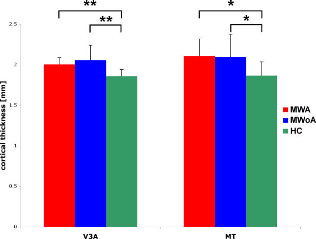
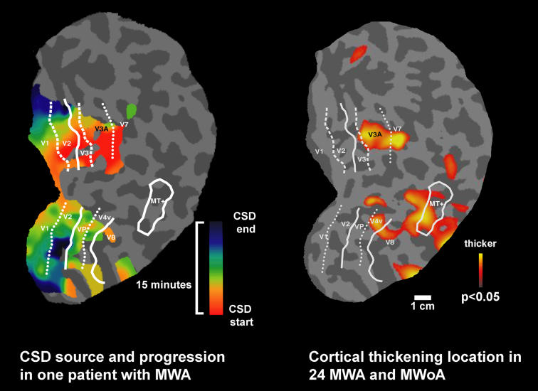
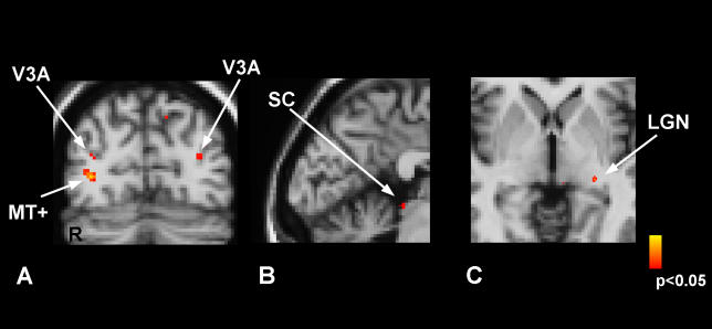
Methods and findings: To investigate a common potential anatomical basis for these disturbances, we used high-resolution cortical thickness measurement and diffusion tensor imaging (DTI) to examine the motion-processing network in 24 migraine patients (12 with MWA and 12 MWoA) and 15 age-matched healthy controls (HCs). We found increased cortical thickness of motion-processing visual areas MT+ and V3A in migraineurs compared to HCs. Cortical thickness increases were accompanied by abnormalities of the subjacent white matter. In addition, DTI revealed that migraineurs have alterations in superior colliculus and the lateral geniculate nucleus, which are also involved in visual processing.
Conclusions: A structural abnormality in the network of motion-processing areas could account for, or be the result of, the cortical hyperexcitability observed in migraineurs. The finding in patients with both MWA and MWoA of thickness abnormalities in area V3A, previously described as a source in spreading changes involved in visual aura, raises the question as to whether a "silent" cortical spreading depression develops as well in MWoA. In addition, these experimental data may provide clinicians and researchers with a noninvasively acquirable migraine biomarker.
Conflict of interest statement
Figures
Comment in
-
The migrainous brain: what you see is not all you get?PLoS Med. 2006 Oct;3(10):e404. doi: 10.1371/journal.pmed.0030404. PLoS Med. 2006. PMID: 17048980 Free PMC article. Review.
References
-
- Lipton RB, Stewart WF. Migraine headaches: Epidemiology and comorbidity. Clin Neurosci. 1998;5:2–9. - PubMed
-
- Roncolato M, Fabbri L, Recchia G, Cavazzuti L, Visona G, et al. An epidemiological study to assess migraine prevalence in a sample of Italian population presenting to their GPs. Eur Neurol. 2000;43:102–106. - PubMed
-
- Warshaw LJ, Burton WN. Cutting the costs of migraine: Role of the employee health unit. J Occup Environ Med. 1998;40:943–953. - PubMed
-
- Stewart WF, Lipton RB, Celentano DD, Reed ML. Prevalence of migraine headache in the United States. Relation to age, income, race, and other sociodemographic factors. JAMA. 1992;267:64–69. - PubMed
-
- Mulleners WM, Chronicle EP, Palmer JE, Koehler PJ, Vredeveld JW. Visual cortex excitability in migraine with and without aura. Headache. 2001;41:565–572. - PubMed
Publication types
MeSH terms
Grants and funding
LinkOut - more resources
Full Text Sources
Miscellaneous
