. 2006 Oct 18:3:36.
doi: 10.1186/1477-7800-3-36.

The role of endoscopic ultrasound in the evaluation of rectal cancer

Affiliations

The role of endoscopic ultrasound in the evaluation of rectal cancer

Ali A Siddiqui et al. Int Semin Surg Oncol. .

Abstract

Accurate staging of rectal cancer is essential for selecting patients who can undergo sphincter-preserving surgery. It may also identify patients who could benefit from neoadjuvant therapy. Clinical staging is usually accomplished using a combination of physical examination, CT scanning, MRI and endoscopic ultrasound (EUS). Transrectal EUS is increasingly being used for locoregional staging of rectal cancer. The accuracy of EUS for the T staging of rectal carcinoma ranges from 80-95% compared with CT (65-75%) and MR imaging (75-85%). In comparison to CT, EUS can potentially upstage patients, making them eligible for neoadjuvant treatment. The accuracy to determine metastatic nodal involvement by EUS is approximately 70-75% compared with CT (55-65%) and MR imaging (60-70%). EUS guided FNA may be beneficial in patients who appear to have early T stage disease and suspicious peri-iliac lymphadenopathy to exclude metastatic disease.


Figures

Figure 1
Figure 1
Correlation between the standard five EUS layers and histological layers of the normal intestinal wall. 1st = interface between fluid in the lumen and the superficial mucosa; 2nd = lamina propria and muscularis mucosa, or deep mucosa; 3rd = submucosa and interface between submucosa and muscularis propria; 4th = muscularis propria; circular (4a) and longitudinal (4c) are not usually seen as separate layers; 5th = interface between serosa and surrounding adventitial tissue.
Figure 2
Figure 2
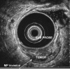
EUS image of T1 rectal cancer confined to mucosa and superficial submucosa. SM=submucosa, MP=muscularis propria, SR=serosa.
Figure 3
Figure 3
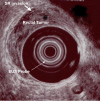
EUS image of T2 rectal cancer invading the muscularis propria.
Figure 4
Figure 4
EUS image of T4 rectal cancer with a metastasis to a perirectal lymph node (L).

References

    1. Jemal A, Siegel R, Ward E, Murray T, Xu J, Smigal C, Thun MJ. Cancer statistics, 2006. CA Cancer J Clin. 2006;56:106–130. - PubMed
    1. Blumberg D, Paty PB, Guillem JG, Picon AI, Minsky BD, Wong WD, Cohen AM. All patients with small intramural rectal cancers are at risk for lymph node metastasis. Dis Colon Rectum. 1999;42:881–885. doi: 10.1007/BF02237095. - DOI - PubMed
    1. Crane CH, Skibber J. Preoperative chemoradiation for locally advanced rectal cancer: rationale, technique, and results of treatment. Semin Surg Oncol. 2003;21:265–270. doi: 10.1002/ssu.10046. - DOI - PubMed
    1. Minsky BD. Adjuvant therapy for rectal cancer--a good first step. N Engl J Med. 1997;336:1016–1017. doi: 10.1056/NEJM199704033361410. - DOI - PubMed
    1. Grann A, Feng C, Wong D, Saltz L, Paty PP, Guillem JG, Cohen AM, Minsky BD. Preoperative combined modality therapy for clinically resectable uT3 rectal adenocarcinoma. Int J Radiat Oncol Biol Phys. 2001;49:987–995. doi: 10.1016/S0360-3016(00)01529-7. - DOI - PubMed

LinkOut - more resources