. 2007 Jan;81(2):783-90.
doi: 10.1128/JVI.01437-06. Epub 2006 Oct 18.

Viral infection of the lungs through the eye

Affiliations

Viral infection of the lungs through the eye

Vira Bitko et al. J Virol. 2007 Jan.

Abstract

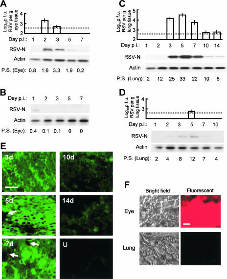
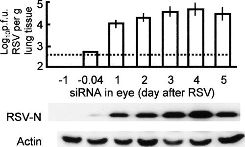
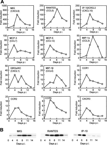
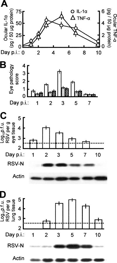
Respiratory syncytial virus (RSV) is the foremost respiratory pathogen in newborns and claims millions of lives annually. However, there has been no methodical study of the pathway(s) of entry of RSV or its interaction with nonrespiratory tissues. We and others have recently established a significant association between allergic conjunctivitis and the presence of RSV in the eye. Here we adopt a BALB/c mouse model and demonstrate that when instilled in the live murine eye, RSV not only replicated robustly in the eye but also migrated to the lung and produced a respiratory disease that is indistinguishable from the standard, nasally acquired RSV disease. Ocularly applied synthetic anti-RSV small interfering RNA prevented infection of the eye as well as the lung. RSV infection of the eye activated a plethora of ocular cytokines and chemokines with profound relevance to inflammation of the eye. Anticytokine treatments in the eye reduced ocular inflammation but had no effect on viral growth in both eye and lung, demonstrating a role of the cytokine response in ocular pathology. These results establish the eye as a major gateway of respiratory infection and a respiratory virus as a bona fide eye pathogen, thus offering novel intervention and treatment options.


Figures

FIG. 1.
FIG. 1.
Replication of ocularly inoculated RSV in the eye and the lung and inhibition by siRNA. RSV was inoculated in BALB/c mouse eyes at day 0. Eye and lung tissues were collected at the indicated days postinoculation (p.i.), and infectious viral titer and viral protein synthesis were determined. The dotted horizontal lines are the lower limits of detection due to serial dilution. (A) RSV growth in the eye. Upper panel, infectious viral titer; lower panel, Western blotting for viral N protein and control cellular actin. P.S., pathology score determined by slit lamp examination (see Materials and Methods). (B) As in panel A, with anti-RSV siRNA administered in the eye 30 min before the virus (2 × 10⁶ PFU per eye). Due to undetectable viral titer, only the Western blot is shown for viral growth. Note the inhibition of the virus, going from panel A to panel B. (C) As in panel A, except that the tissue is lung. (D) Studies of the lung tissue when RSV and anti-RSV siRNA were administered in the eye. All errors bars (standard deviations) are from three experiments. Note that the virus disappears in the eye (A) as it appears in the lung (B), suggesting travel. Also, compare panel A with B and panel C with D to note the drastic inhibition of the virus by siRNA. (E) Immunostaining for RSV in lung sections on different days after RSV infection. U, uninfected (i.e., no virus was put in the eye). The arrows point to vascular spaces. Bar, 400 μm. (F) Bright-field and fluorescent images of eye and lung sections at 24 h after instillation of Cy3-labeled siRNA in the eye. Bar, 400 μm. Brain and liver sections looked essentially like those of the lung (i.e., lacked fluorescence) and hence are not shown.
FIG. 2.
FIG. 2.
Effect of siRNA administered postinfection. Mouse experiments were performed essentially as for Fig. 1, except that the siRNA was administered in the eye at the following times relative to RSV (x axis, from left): 1 day before RSV; 0.04 day (1 h) before RSV; and then 1, 2, 3, 4, or 5 days after RSV. For titer and protein analyses, lungs were isolated at 5 days after RSV infection because this time point showed maximal titer (e.g., see Fig. 1). Error bars indicate standard deviations. The lower panel shows immunoblots for virus growth (RSV N protein) and an actin control for the same tissue samples.
FIG. 3.
FIG. 3.
Induction of cytokines and receptors in RSV-infected eye. (A) mRNA profiles. The RNAs isolated from the RSV-infected (and uninfected control) eyes at different days postinfection (p.i.) were subjected to quantitative real-time RT-PCR as described in Materials and Methods. The relative amounts of RNA were expressed as the ratio to the uninfected control value (fold induction). Each box represents a specific cytokine, with its common name given and the uniform ligand name in parentheses. Each data point is derived from three independent infection experiments; the errors were less than 15% and are omitted for simplicity. (B) Protein profiles of the top three representative cytokines detected by Western blotting at the same time points as the RNA. Actin was present in equal amounts in all lanes (not shown to save space).
FIG. 4.
FIG. 4.
Anticytokine treatment of the RSV-infected eye and its effect. Infection and treatment are described in Materials and Methods. (A) IL-1α and TNF-α levels in the eye assayed by ELISA at different days postinfection (p.i.). (B) Pathology score of the eye and its reduction by neutralizing antibodies against IL-1α and TNF-α (white bars, untreated; stippled bars, IL-1α; hatched bars, TNF-α). The scores are also numerically presented in Fig. 1A. (C and D) RSV growth in the eye and the lung, respectively, when anti-IL-1α antibody was administered in the eye along with RSV. Upper panel, infectious RSV titer; lower panel, Western blot for viral N protein and control cellular actin. Note the relatively greater RSV replication in panels C and D compared to the untreated tissues in panels A and B of Fig. 1. All error bars (standard deviations) are derived from three experiments. In all antibody experiments, nonimmune mouse IgG was used as a control in at least three eyes, and no pathology was seen.

References

    1. Adamus, G., M. Manczak, and M. Machnicki. 2001. Expression of CC chemokines and their receptors in the eye in autoimmune anterior uveitis associated with EAE. Investig. Ophthalmol. Vis. Sci. 42:2894-2903. - PubMed
    1. Barik, S. 2004. Control of nonsegmented negative-strand RNA virus replication by siRNA. Virus Res. 102:27-35. - PubMed
    1. Barik, S. 2005. Silence of the transcripts: RNA interference in medicine. J. Mol. Med. 83:764-773. - PubMed
    1. Bitko, V., and S. Barik. 2001. Phenotypic silencing of cytoplasmic genes using sequence-specific double-stranded short interfering RNA and its application in the reverse genetics of wild type negative-strand RNA viruses. BMC Microbiol. 1:34. - PMC - PubMed
    1. Bitko, V., N. E. Garmon, T. Cao, B. Estrada, J. E. Oakes, R. N. Lausch, and S. Barik. 2004. Activation of cytokines and NF-kappa B in corneal epithelial cells infected by respiratory syncytial virus: potential relevance in ocular inflammation and respiratory infection. BMC Microbiol. 4:28. - PMC - PubMed

Publication types

MeSH terms