. 2007 Jan;81(1):280-91.
doi: 10.1128/JVI.01616-06. Epub 2006 Oct 18.

Glycoprotein L disruption reveals two functional forms of the murine gammaherpesvirus 68 glycoprotein H

Affiliations

Glycoprotein L disruption reveals two functional forms of the murine gammaherpesvirus 68 glycoprotein H

Laurent Gillet et al. J Virol. 2007 Jan.

Abstract

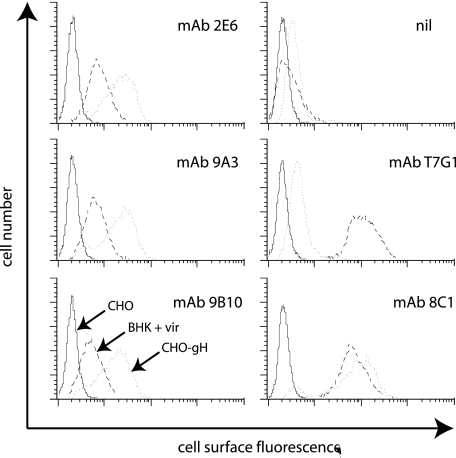
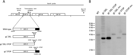
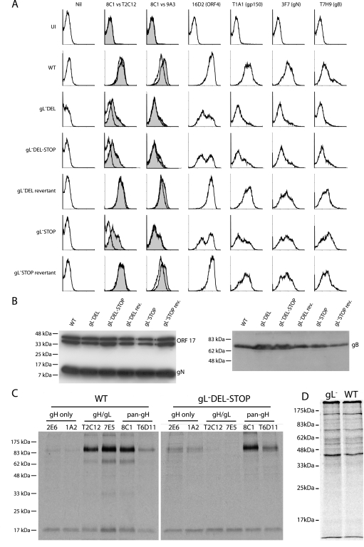
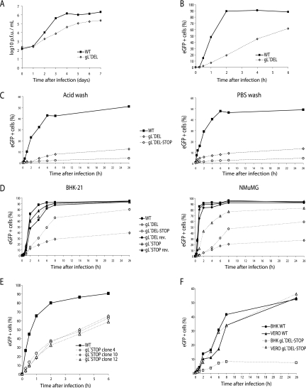
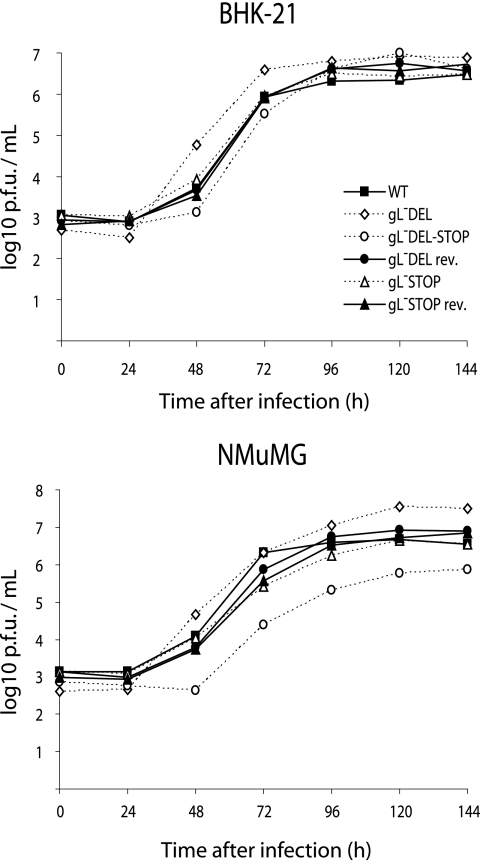
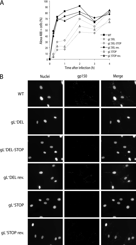
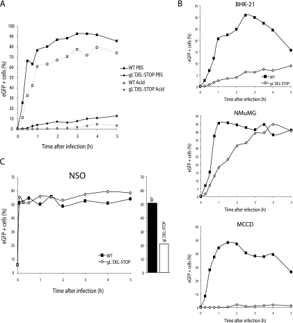
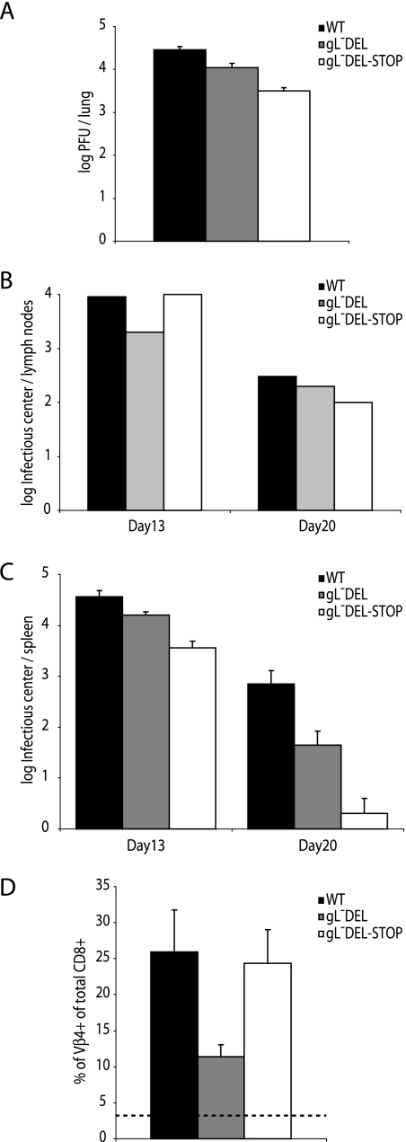
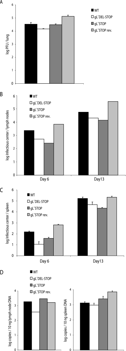
The herpesvirus glycoprotein H (gH) and gL associate to form a heterodimer that plays a central role in virus-driven membrane fusion. When archetypal alpha- or betaherpesviruses lack gL, gH misfolds and progeny virions are noninfectious. In order to define the role that gL plays in gamma-2 herpesvirus infections, we disrupted its coding sequence in murine gammaherpesvirus-68 (MHV-68). MHV-68 lacking gL folded gH into a conformation antigenically distinct from the form that normally predominates on infected cells. gL-deficient virions bound less well than the wild type to epithelial cells and fibroblasts. However, they still incorporated gH and remained infectious. The cell-to-cell spread of gL-deficient viruses was remarkably normal, as was infection, dissemination, and latency establishment in vivo. Viral membrane fusion was therefore gL independent. The major function of gL appeared to be allowing gH to participate in cell binding prior to membrane fusion. This function was most important for the entry of MHV-68 virions into fibroblasts and epithelial cells.


Figures

FIG. 1.
FIG. 1.
Flow-cytometric identification of gL-dependent and gL-independent MAbs recognizing the MHV-68 gH. BHK-21 cells were infected (18 h, 2 PFU/cell) with wild-type MHV-68 (BHK + vir). CHO-gH cells stably express a glycosylphosphatidylinositol-linked form of the gH extracellular domain without gL. MAbs 2E6, 9A3, and 9B10 were selected for their recognition of CHO-gH cells. MAb T7G1 recognizes gH on MHV-68-infected cells but not on cells expressing only gH. MAb 8C1 recognizes gH in either context. nil, secondary antibody only.
FIG. 2.
FIG. 2.
Generation of gL-deficient MHV-68 mutants. A. The gL coding sequence within ORF47 was disrupted by deleting its 5′ end including its start codon (gL-DEL), by combining the deletion with premature stop codons (gL-DEL-STOP), or by inserting stop codons near the end of the coding sequence for its predicted signal peptide (gL-STOP). Each mutation incorporated a new BamHI restriction site. B. Viral DNA was digested with BamHI, resolved by agarose gel electrophoresis, and hybridized with a 32P-labeled BamHI-N probe, as shown in panel A. The 4,048-bp wild-type (WT) band becomes 3,375 bp for the gL-DEL and gL-DEL-STOP mutants and 3,433-bp for the gL-STOP mutant. The remaining 600-bp fragments are too small to appear on this gel.
FIG. 3.
FIG. 3.
Glycoprotein expression by gL-deficient MHV-68. A. BHK-21 cells were left uninfected (UI) or infected (1 PFU, 24 to 48 h) with gL or gL+ viruses as indicated. The cells were then trypsinized and stained for viral glycoproteins. nil, secondary antibody only. For gH expression, gH/gL-specific (T2C12) and gH-only (9A3) MAbs (open histograms) are shown in comparison to a pan-gH MAb (8C1) (filled histograms). eGFP-expressing viruses were used, and each histogram shows an equivalent eGFP+-gated population to ensure equivalent levels of infection. WT, wild type. B. Viral proteins (equivalent to 5 × 10⁴ PFU/lane for each virus) were denatured, resolved by SDS-PAGE, and immunoblotted for gB and gN as indicated. MAb 7D1-C12 specifically detects ORF17, either in cells transfected with an ORF17 expression plasmid or as a glutathione S-transferase-ORF17 fusion protein expressed in Escherichia coli (data not shown). It provides a loading control. The doublet band reflects the fact that ORF17 is autoproteolytically cleaved. C. gH was immunoprecipitated from 35S-labeled virions. The MAbs used were gH only (2E6, 1A2), gH/gL specific (T2C12, 7E5), or pan-gH (8C1, T6D11). gH migrates at an apparent molecular mass of 85 kDa. The nonspecifically immunoprecipitated 19-kDa band probably corresponds to the abundant ORF65 capsid component. D. Input labeled virion proteins used for immunoprecipitation in panel C.
FIG. 4.
FIG. 4.
Infectivity assays with gL-deficient MHV-68 mutants. A. BHK-21 cells were infected (0.01 PFU/cell, 1 h, 37°C) with wild-type (WT) or gL-DEL MHV-68. Infectious virus in replicate cultures was determined thereafter by plaque assay. B. BHK-21 cells were exposed to eGFP-expressing wild-type or gL-DEL MHV-68 (1 PFU/cell) for the times indicated and then washed with PBS and cultured overnight. Viral infection was assayed by flow cytometry for eGFP expression. C. MEFs were exposed to eGFP-expressing wild-type, gL-DEL, or gL-DEL-STOP MHV-68 (1 PFU/cell) for the times indicated and then washed either with PBS (pH 7.4) or with isotonic (pH 3) buffer (acid wash). Viral infection was assayed by measuring eGFP expression 18 h later as for panel B. D. BHK-21 fibroblasts or NMuMG epithelial cells were infected with eGFP-expressing gL or gL+ viruses as for panel B. The cells were washed with PBS after the time indicated, and eGFP expression was assayed the next day. E. Three different BAC isolates of the gL-STOP mutant were reconstituted into infectious virus and then compared with the wild type for their capacity to infect BHK-21 cells. F. BHK-21 and Vero cells were exposed to gL or gL+ MHV-68 for different times before being washed with PBS as for panel D.
FIG. 5.
FIG. 5.
Growth of gL-deficient MHV-68 with allowance made for reduced cell binding. BHK-21 fibroblasts or NMuMG fibroblasts were infected at low multiplicity (0.01 PFU/cell) with gL or gL+ MHV-68 and then cultured without removing the virus inoculum. The infectious virus in replicate cultures was determined thereafter by plaque assay. WT, wild type.
FIG. 6.
FIG. 6.
Decreased cell binding by gL-deficient MHV-68 mutants. A. BHK-21 cells were exposed to gL or gL+ viruses (2 PFU/cell) at 37°C for the times indicated and then fixed, permeabilized, and stained with the gN-specific MAb 3F7 plus an Alexa 488-conjugated mouse IgG-specific secondary antibody. Cells were analyzed for positive staining by flow cytometry. WT, wild type. B. BHK-21 cells were exposed to gL or gL+ viruses for 30 min as in panel A and then washed with PBS and stained for gp150 with MAb LSB11 plus an Alexa 488-conjugated secondary antibody. Nuclei were counterstained with DAPI.
FIG. 7.
FIG. 7.
Tracking cell binding with eGFP-tagged MHV-68. A. BHK-21 cells were exposed to either wild-type (WT) or gL-deficient versions of MHV-68 with eGFP-tagged gM (2 PFU/cell) and then cultured at 37°C. The cells were washed with PBS or at low pH after the time indicated and analyzed by flow cytometry for green fluorescence. B. BHK-21 fibroblasts, NMuMG epithelial cells, and MCCD epithelial cells were exposed to gL or gL+ versions of the gM-eGFP virus as for panel A. The cells were washed with PBS at the time indicated and then analyzed by flow cytometry for green fluorescence. The decline from maximum fluorescence with time may reflect the destruction of gM-eGFP in lysosomes following endocytosis and fusion. C. NS0 cells were analyzed for green fluorescence after exposure to gL or gL+ gM-eGFP MHV-68 as for panel C. A 2-h parallel infection of BHK-21 cells at the same multiplicity (2 PFU/cell) is shown.
FIG. 8.
FIG. 8.
Replication of gL and gL+ MHV-68 in C57BL/6 mice. A. C57BL/6 mice were infected intranasally with gL+ or gL viruses. Seven days later, the infectious virus titer in lungs was determined by plaque assay. The titers of gL viruses were significantly reduced relative to the wild type (WT) (P < 0.02 by Student's t test). B. Mediastinal lymph nodes were removed at the times indicated after infection with gL+ or gL viruses, pooled from five mice, and assayed for reactivable MHV-68 by infectious-center assay. C. Spleens from the same mice were analyzed individually by infectious-center assay. Each bar shows the mean ± standard error of the mean (SEM) for each group of five. The gL-DEL titers were not significantly different from the wild type at day 13. The other gL virus titers did show a significant reduction (P < 0.03). D. Splenic CD8+ T cells were analyzed for Vβ4+ subset expansion at 20 days postinfection. Each bar shows the mean ± SEM for five mice per group. The dashed line shows the value for naive mice.
FIG. 9.
FIG. 9.
Replication of gL and gL+ MHV-68 in BALB/c mice. A. BALB/c mice were infected intranasally with gL+ or gL viruses. Six days later, the infectious virus titer in lungs was determined by plaque assay. There was no significant difference between gL and wild-type (WT) virus titers. B. Mediastinal lymph nodes were removed at the times indicated after infection with gL+ or gL viruses, pooled from five mice, and assayed for reactivable MHV-68 by infectious-center assay. C. Spleens from the same mice were analyzed individually by infectious-center assay. Each bar shows the mean ± standard error of the mean (SEM) for each group of five. At day 6, the gL titers were significantly lower than wild type (P < 0.01 by Student's t test). By day 13, only the gL-DEL-STOP titers were lower (P < 0.03). D. DNA was extracted from individual spleens. The viral genome copy number was then determined by real-time PCR amplification of genomic coordinates 24832 to 25071 and comparison with parallel amplifications of plasmid template dilutions. Mediastinal lymph nodes were assayed as pools of five mice, spleens were assayed individually, and each bar shows the mean ± SEM. The gL viruses were not significantly different from the wild type.

References

    1. Adler, H., M. Messerle, M. Wagner, and U. H. Koszinowski. 2000. Cloning and mutagenesis of the murine gammaherpesvirus 68 genome as an infectious bacterial artificial chromosome. J. Virol. 74:6964-6974. - PMC - PubMed
    1. Bennett, N. J., J. S. May, and P. G. Stevenson. 2005. Gamma-herpesvirus latency requires T cell evasion during episome maintenance. PLoS Biol. 3:e120. - PMC - PubMed
    1. Blaskovic, D., M. Stancekova, J. Svobodova, and J. Mistrikova. 1980. Isolation of five strains of herpesviruses from two species of free living small rodents. Acta Virol. 24:468. - PubMed
    1. Boname, J. M., and P. G. Stevenson. 2001. MHC class I ubiquitination by a viral PHD/LAP finger protein. Immunity 15:627-636. - PubMed
    1. Boname, J. M., B. D. de Lima, P. J. Lehner, and P. G. Stevenson. 2004. Viral degradation of the MHC class I peptide loading complex. Immunity 20:305-317. - PubMed

Publication types

MeSH terms

LinkOut - more resources