. 2006 Oct 31;103(44):16129-34.
doi: 10.1073/pnas.0607467103. Epub 2006 Oct 18.

Three strategically placed hydrogen-bonding residues convert a proton pump into a sensory receptor

Affiliations

Three strategically placed hydrogen-bonding residues convert a proton pump into a sensory receptor

Yuki Sudo et al. Proc Natl Acad Sci U S A. .

Abstract

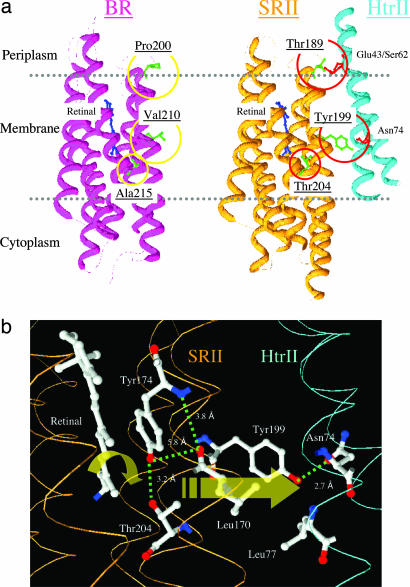
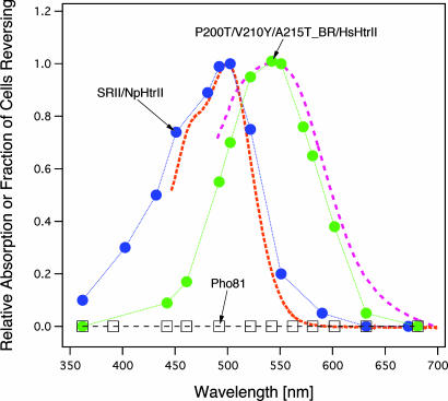
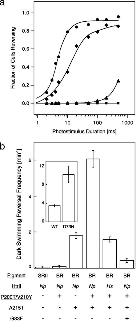
In haloarchaea, light-driven ion transporters have been modified by evolution to produce sensory receptors that relay light signals to transducer proteins controlling motility behavior. The proton pump bacteriorhodopsin and the phototaxis receptor sensory rhodopsin II (SRII) differ by 74% of their residues, with nearly all conserved residues within the photoreactive retinal-binding pocket in the membrane-embedded center of the proteins. Here, we show that three residues in bacteriorhodopsin replaced by the corresponding residues in SRII enable bacteriorhodopsin to efficiently relay the retinal photoisomerization signal to the SRII integral membrane transducer (HtrII) and induce robust phototaxis responses. A single replacement (Ala-215-Thr), bridging the retinal and the membrane-embedded surface, confers weak phototaxis signaling activity, and the additional two (surface substitutions Pro-200-Thr and Val-210-Tyr), expected to align bacteriorhodopsin and HtrII in similar juxtaposition as SRII and HtrII, greatly enhance the signaling. In SRII, the three residues form a chain of hydrogen bonds from the retinal's photoisomerized C(13)=C(14) double bond to residues in the membrane-embedded alpha-helices of HtrII. The results suggest a chemical mechanism for signaling that entails initial storage of energy of photoisomerization in SRII's hydrogen bond between Tyr-174, which is in contact with the retinal, and Thr-204, which borders residues on the SRII surface in contact with HtrII, followed by transfer of this chemical energy to drive structural transitions in the transducer helices. The results demonstrate that evolution accomplished an elegant but simple conversion: The essential differences between transport and signaling proteins in the rhodopsin family are far less than previously imagined.


Conflict of interest statement

The authors declare no conflict of interest.

Figures

Fig. 1.
Fig. 1.
Locations of residues in this study. (a) X-ray crystal structures of BR (PDB ID code 1C3W) and SRII/HtrII (PDB ID code 1H2S) complex in the dark, indicating the three hydrogen-bonding residues in SRII introduced into BR in this study: Pro-200, Val-210, and Ala-215 in BR that correspond to Thr-189, Tyr-199, and Thr-204 in SRII, respectively. Tyr-199 bonds with HtrII Asn-74; Thr-189 bonds with HtrII Glu-43 and Ser-62; and Thr-204 with retinal pocket residue Tyr-174, which is conserved in BR as Tyr-185. (b) Detail of the x-ray SRII/HtrII structure (16), which focuses on the midmembrane SRII–HtrII interface containing the core signal relay structure identified in this work.
Fig. 2.
Fig. 2.
Photoinduced proton transport of WT BR (a) and BR mutant P200T/V210Y (b) with and without HtrII. (a) Pho81Wr vesicles not expressing BR are included as a negative control. We measured proton pumping activity of BR (WT and P200T/V210Y mutant) with and without HtrII. Light-driven proton transport by BR mutant P200T/V210Y is ceased by association with the transducer protein, HtrII. We infer that association with HtrII closes a cytoplasmic channel of BR as it does in SRI (12).
Fig. 3.
Fig. 3.
Flash-induced absorption changes of WT BR with (blue) and without (red) HtrII (Upper) and BR mutant P200T/V210Y with (blue) and without (red) HtrII (Lower). Scale in absorption units (ordinate) and time (abscissa). Note the 20-fold difference in the time scales. The M intermediate is monitored by 410 nm and the unphotolysed state is monitored by 570 nm. Flash photolysis data were acquired from membrane samples in 4 M NaCl at pH 7.0 in transformants of H. salinarum strain Pho81Wr, which lacks rhodopsins and the transducer proteins HtrI and HtrII.
Fig. 4.
Fig. 4.
Phototaxis responses of BR–HtrII complexes. Swimming reversal frequency responses of cell populations measured by stimulus (at dotted arrow) effects on the ratio of rate of change of direction (RCD) to speed (SPD) by computer-assisted motion analysis. There were 200–2,500 cells assayed for each trace. “BR” designates BR in the following description: 100-ms 500-nm stimulus for SRII/HtrII (a), 500-ms 580-nm stimulus for P200T/V210Y-BR (b), 500-ms 550-nm stimulus for A215T-BR (c), P200T/V210Y/A215T-BR/NpHtrII (d), and P200T/V210Y/A215T-BR/G83F-NpHtrII (j), and 100-ms 550-nm stimulus for P200T/V210Y/A215T-BR/HsHtrII (e). BR mutants containing the A215T mutation have shorter absorbance maxima (λmax, 550 nm) than WT BR (λmax, 580 nm) (27). Swimming reversal frequency transients to a step-down in continuous light as indicated (f–i%). (f) SRII/HtrII; (g) P200T/V210Y/A215T-BR/NpHtrII; (h) P200T/V210Y/A215T-BR/HsHtrII; and (i) P200T/V210Y/A215T-BR/G83F-NpHtrII.
Fig. 5.
Fig. 5.
Action spectra for the repellent response in transformants containing SRII/NpHtrII (blue circles), BR triple mutant/HsHtrII (green circles), and Pho-81 devoid of rhodopsins (squares). The absorption spectra (dotted lines) were measured by using the membrane fraction in the presence of 0.1% n-dodecyl-β-d-maltoside (26). We measured phototaxis responses as swimming reversal frequency changes to a 5-ms photostimulus for SRII/NpHtrII and 10-ms photostimuli for the BR triple mutant/HsHtrII and Pho-81.
Fig. 6.
Fig. 6.
Photostimulus-induced and dark swimming reversal frequencies. (a) Fluence/response curves of cells containing SRII/HtrII (circles), BR triple mutant/HsHtrII (diamonds), BR triple mutant/NpHtrII (triangles), and WT BR/NpHtrII (squares). Km values from Michaelis–Menton fits to the fluence/response curves were used to estimate sensitivities of cells expressing BR triple mutant/HsHtrII and BR triple mutant/NpHtrII (35% and 0.4%, respectively, of that conferred by SRII-HtrII). (b) Dark swimming reversal frequency of cells expressing WT and mutated BR and SRII. Increase in frequency corresponds to constitutive activity in the dark (37). The values were determined from duplicate measurements of ≈30 cells imaged by infrared illumination and tracked for 2 min. (b Inset) Dark reversal frequencies of cells containing the WT and D73N mutant of H. salinarum SRII–HtrII complexes studied in ref. .

References

    1. Ruiz-Gonzalez MX, Marin I. J Mol Evol. 2004;58:348–358. - PubMed
    1. Spudich JL, Jung K-H. In: Handbook of Photosensory Receptors. Briggs W, Spudich JL, editors. Weinheim, Germany: Wiley; 2005. pp. 1–24.
    1. Sharma AK, Spudich JL, Doolittle WF. Trends Microbiol. 2006 Sep 26; 10.1016/j.tim.2006.09.006. - PubMed
    1. Luecke H, Lanyi JK. Adv Protein Chem. 2003;63:111–130. - PubMed
    1. Béjà O., Spudich EN, Spudich JL, Leclerc M, DeLong EF. Nature. 2001;411:786–789. - PubMed

Publication types

LinkOut - more resources