. 2006 Oct 31;103(44):16278-83.
doi: 10.1073/pnas.0607210103. Epub 2006 Oct 18.

Macrophage inhibitory cytokine 1 mediates a p53-dependent protective arrest in S phase in response to starvation for DNA precursors

Affiliations

Macrophage inhibitory cytokine 1 mediates a p53-dependent protective arrest in S phase in response to starvation for DNA precursors

Mukesh K Agarwal et al. Proc Natl Acad Sci U S A. .

Abstract

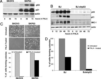
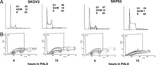
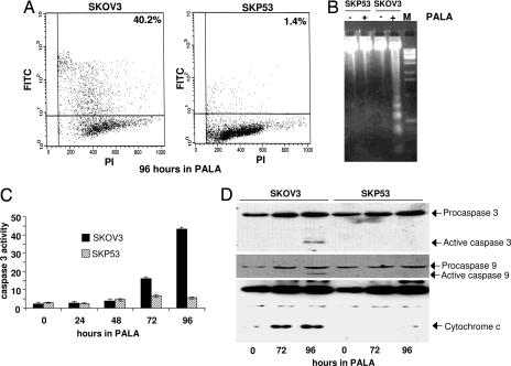
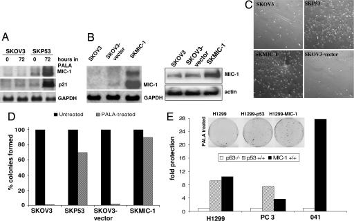
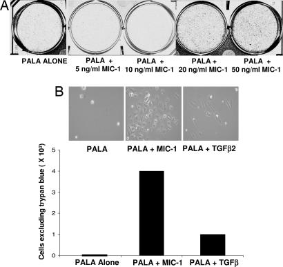
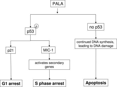
p53 is essential for the cellular responses to DNA damage that help to maintain genomic stability. Protective p53-dependent cell-cycle checkpoints are activated in response to a wide variety of stresses, including not only DNA damage but also arrest of DNA synthesis and of mitosis. In addition to its role in activating the G(1) and G(2) checkpoints, p53 also helps to protect cells in S phase when they are starved for DNA precursors by treatment with the specific aspartate transcarbamylase inhibitor N-phosphonacetyl-l-aspartate (PALA), which blocks the synthesis of pyrimidine nucleotides. Even though p53 is activated, PALA-treated cells expressing low levels of p53 or lacking expression of p21 do not arrest in G(1) or G(2) but are blocked in S phase instead. In the complete absence of p53, PALA-treated cells continue to synthesize DNA slowly and eventually progress through S phase, suffering severe DNA damage that in turn triggers apoptosis. Expression of the secreted protein macrophage inhibitory cytokine 1 (MIC-1), a member of the TGF-beta superfamily, increases substantially after PALA treatment, and application of exogenous MIC-1 or its constitutive expression from a cDNA provides remarkable protection of p53-null cells from PALA-mediated apoptosis, arguing that the p53-dependent secretion of MIC-1 provides a major part of such protection. Stimulation of MIC-1-dependent S phase arrest in normal gut epithelial cells might help to revitalize the clinical use of PALA, which has been limited by gut toxicity.


Conflict of interest statement

The authors declare no conflict of interest.

Figures

Fig. 1.
Fig. 1.
p53 protects PALA-treated cells. Cells were treated with 250 μM PALA for 12–72 h. (A and B) Expression of p53, p21, and MIC-1, analyzed by the Western blot method. (C and D) Colony formation for cells treated with 250 μM PALA for 12 days. Cells plated for colony formation were assayed after an additional 15 days.
Fig. 2.
Fig. 2.
Cells expressing p53 stop making DNA in PALA, whereas those lacking p53 continue to cycle. (A) FACS analysis of SKOV3 and SKP53 cells treated with 250 μM PALA for 72 h. The cells were stained with propidium iodide to measure DNA content. Percentages of cells in G1, G2/M, and S are shown in each image. (B) Incorporation of BrdU. Untreated and treated SKOV3 and SKP53 cells were labeled with BrdU for 4 h, fixed in methanol, stained with anti-BrdU, and measured by FACS.
Fig. 3.
Fig. 3.
Cells lacking p53 undergo apoptosis in PALA. SKOV3 and SKP53 cells were treated with 250 μM PALA for the times indicated. (A) Fraction of apoptotic cells, determined by the TUNEL assay. (B) DNA laddering in untreated and PALA-treated SKOV3 and SKP53 cells. (C) Activation of caspase 3 in PALA-treated cells. (D) Activation of caspases 9 and 3 and release of cytochrome c in PALA-treated cells. Proteins were measured by the Western blot method.
Fig. 4.
Fig. 4.
Constitutive expression of MIC-1 protects PALA-treated cells lacking p53. (A) Analysis of MIC-1 and p21 mRNA expression. The Northern blot method was used. (B) Analysis of expression of MIC-1 in cells transfected with MIC-1 cDNA by the Northern (Left) and Western (Right) blot methods. (C) Growth of cells in PALA. SKOV3, SKP53, SKMIC-1, and SKOV3 vector. Cells were treated with 250 μM PALA, and photographs were taken 7 days later. (D) Colony formation in SKOV3, SKP53, and SKMIC-1 cells treated with 250 μM PALA for 12 days. The percentage of colonies formed with individual cloned cell lines is shown. Similar results were obtained with pools (not shown). (E) Colony formation in H1299, PC3, 041, and its counterparts expressing MIC-1 or p53, treated with 250 μM PALA for 12 days. Fold protection was calculated by dividing the number of colonies formed in p53- or MIC-1-expressing cells by the number formed in parental p53-null cells.
Fig. 5.
Fig. 5.
Treatment with MIC-1 protects p53-null SKOV3 cells from PALA. (A) Cells in 250 μM PALA were treated with various concentrations of MIC-1 (replenished daily), and after 12 days, the plates were stained with methylene blue and photographed. (B) Cells were treated with a 50 ng/ml concentration of either MIC-1 or TGF-β2 and 250 μM PALA. Twelve days later, the cells were photographed, and their viability was measured by trypan blue exclusion.
Fig. 6.
Fig. 6.
p53-dependent effects on cell-cycle checkpoints in response to PALA.

References

    1. Linke SP, Clarkin KC, Di Leonardo A, Tsou A, Wahl GM. Genes Dev. 1996;10:934–947. - PubMed
    1. Taylor WR, Agarwal ML, Agarwal A, Stacey DW, Stark GR. Oncogene. 1999;18:283–295. - PubMed
    1. Cross SM, Sanchez CA, Morgan CA, Schimke MK, Ramel S, Idzerda RL, Raskind WH, Reid BJ. Science. 1995;267:1353–1356. - PubMed
    1. Di Leonardo A, Khan SH, Linke SP, Greco V, Seidita G, Wahl GM. Cancer Res. 1997;57:1013–1019. - PubMed
    1. Swyryd EA, Seaver SS, Stark GR. J Biol Chem. 1974;249:6945–6950. - PubMed

Publication types

MeSH terms