Comparative Study
. 2006 Oct 18;26(42):10646-57.
doi: 10.1523/JNEUROSCI.2305-06.2006.

Trigeminal neuropathic pain alters responses in CNS circuits to mechanical (brush) and thermal (cold and heat) stimuli

Affiliations
Comparative Study

Trigeminal neuropathic pain alters responses in CNS circuits to mechanical (brush) and thermal (cold and heat) stimuli

Lino Becerra et al. J Neurosci. .

Abstract

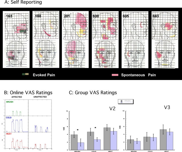
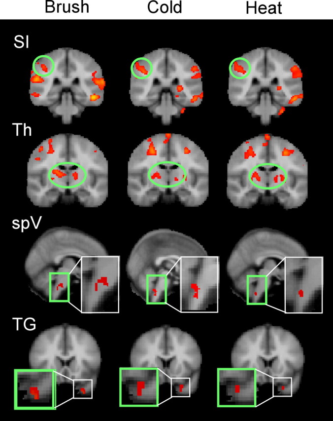
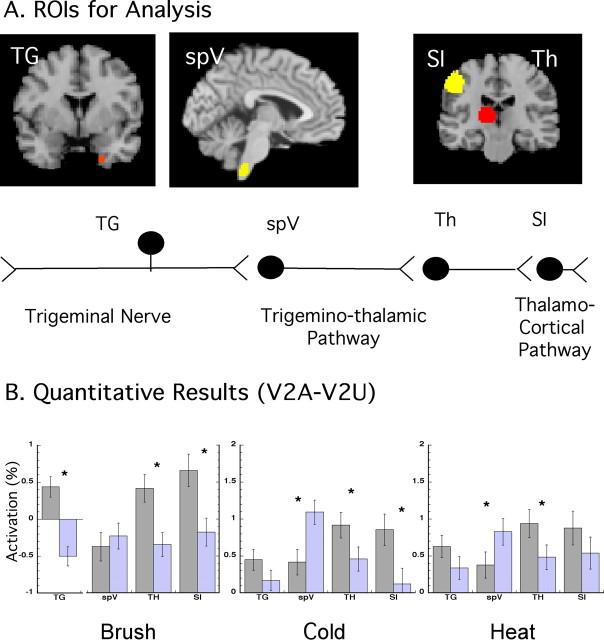
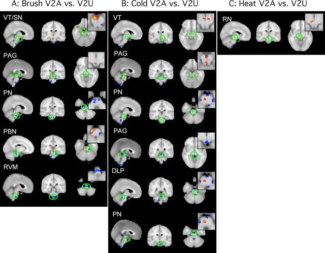
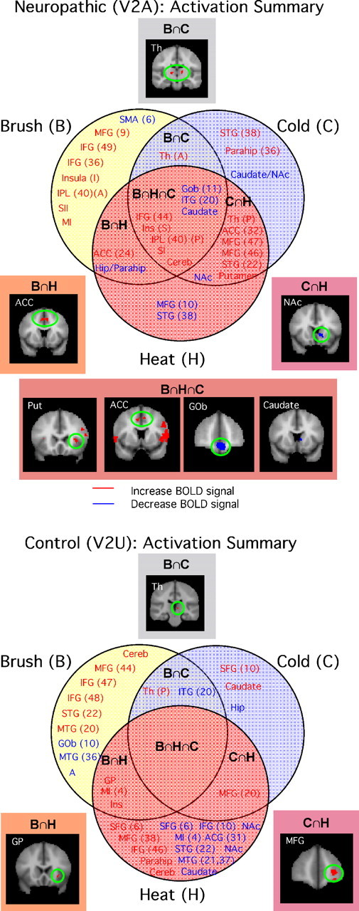
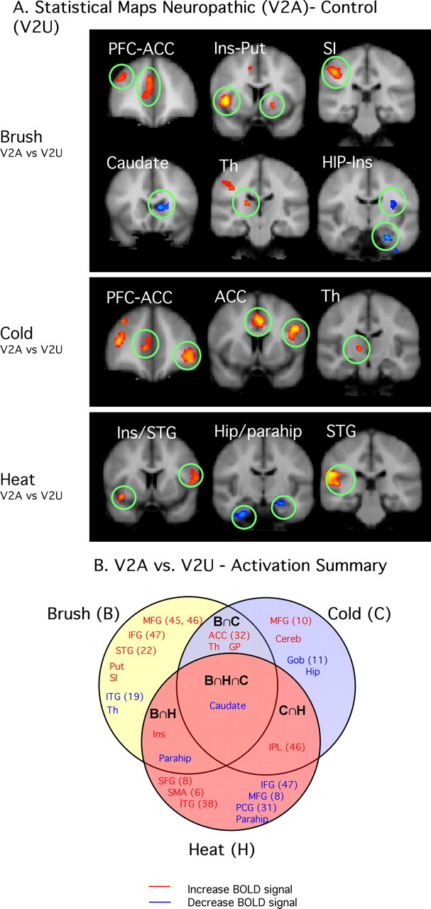
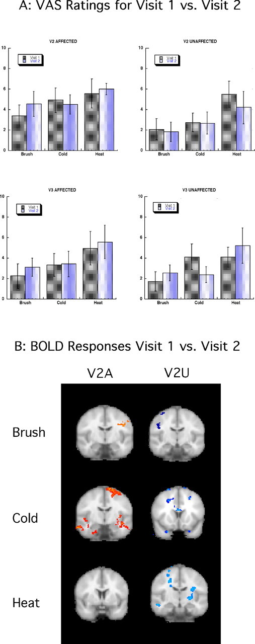
Functional magnetic resonance imaging was used to study patients with chronic neuropathic pain involving the maxillary region (V2) of the trigeminal nerve in patients with spontaneous pain and evoked pain to brush (allodynia). Patients underwent two functional scans (2-3 months apart) with mechanical and thermal stimuli applied to the affected region of V2 and to the mirror site in the unaffected contralateral V2 region, as well as bilaterally to the mandibular (V3) division. Patients were stimulated with brush, noxious cold, and noxious heat. Significant changes were observed in regions within and outside the primary trigeminal sensory pathway. Stimulation to the affected (neuropathic) side resulted in predominantly frontal region and basal ganglia activation compared with the control side. The differences were consistent with the allodynia to brush and cold. A region of interest-based analysis of the trigeminal sensory pathway revealed patterns of activation that differentiated between the affected and unaffected sides and that were particular to each stimulus. Activation in the spinal trigeminal nucleus was constant in location for all pain stimuli. Activation in other brainstem nuclei also showed differences in the blood oxygenation level-dependent signal for the affected versus the unaffected side. Thus, sensory processing in patients with trigeminal neuropathic pain is associated with distinct activation patterns consistent with sensitization within and outside of the primary sensory pathway.


Figures

Figure 1.
Figure 1.
A, Facial maps. Subjects were asked to draw on divided frontal and lateral views of the face the areas in which they experience spontaneous (red) and evoked (yellow/green) pain. This information was used to define the area for stimulation in the scanner. B, VAS on-line ratings. On-line VAS ratings for one subject in response to brush, cold, and heat applied to the affected and unaffected V2 areas are shown. The response to thermal stimuli in the affected area seemed to last longer and produced lingering pain not observed in the ratings of the unaffected area. C, Group results. VAS group results obtained from the on-line ratings for all subjects for V2A versus V2U (left) and all visits to brush, cold, and heat are shown. Brush and cold achieved statistically significant differences between sides (t test, *p < 0.05), but heat did not. V3 affected versus unaffected (right) stimulation produced ratings that were not significantly different to V2 unaffected. Error bars indicate SEM.
Figure 2.
Figure 2.
Group analysis for the trigeminal pathway. Specific activations along the trigeminal pathway (SI, thalamus, spV, and TG) are presented for the three stimuli. SI displayed similar activation across the three stimuli; the thalamus seemed to have a more medial activation with brush compared with heat and cold. Aggregate image analysis of the TG and trigeminal nuclei did not meet statistical threshold. Subthreshold activation was seen for spV in brush, but only spV for heat and cold. The TG activated similarly for the three stimuli. Quantified analysis through an ROI-based analysis is shown in Figure 3. Th, Thalamus.
Figure 3.
Figure 3.
ROI-based analysis for trigeminal pathway. A, ROI-based approach analysis. The location of the ROIs from which z values were extracted to perform an ROI-based analysis (see Materials and Methods) is shown. B, The bar graphs indicate mean differences in percentage signal change (with SEM) for each ROI for the three stimuli. TG was significantly more active in V2A for the three stimuli. The spV was more active in the unaffected area, and the SI and thalamus were more active on V2A than V2U not achieving significance SI for heat. See ROI-based results. Th, Thalamus.
Figure 4.
Figure 4.
Brainstem activation. Brainstem activation differences of V2A versus V2U for the three stimuli are shown. Images are presented in three planes with a zoom-in image for axial slices. A, Activation differences to brush stimulation. B, Activation differences to cold stimulation, C, Activation differences to heat stimulation. Red-yellow, V2A > V2U; blue, V2A < V2U; PBN, parabrachial nucleus; RVM, rostral ventral medulla; DLP, dorsolateral pons.
Figure 5.
Figure 5.
Summary of regions activated by brush, cold, and heat. A summary of the aggregate image results for V2A and V2U according to a Venn diagram is shown. Top, Neuropathic: significant activation was found in common for the three stimuli. Note also that significant commonality between heat and cold stimuli was observed. See Activation in affected and unaffected regions. Bottom, Control: activation in V2U to the three stimuli resulted in very little overlap between stimuli and not all among the three of them. See text. STG, Superior temporal gyrus; IPL, inferior parietal lobe; SII, secondary somatosensory cortex; MI, motor cortex; Th, thalamus; Parahip, parahippocampal gyrus; ITG, inferior temporal gyrus; MTG, medial temporal gyrus; PCG, posterior cingulate cortex; A, amygdala; Hip, hippocampus; Cereb, cerebellum; Put, putamen. Letters in parentheses after structure label: S, superior; I, inferior; A, anterior; P, posterior.
Figure 6.
Figure 6.
Summary of regions showing differences between neuropathic and control sides. A, Contrast maps for the three stimuli applied to the affected area (V2A vs V2U). Brush displayed significant differences in sensory/discriminative and emotional/cognitive areas, whereas cold and heat seem to have main differences in nonsensory pathways. See text. B, Venn diagram depicting commonalities of contrasts for the three stimuli. Caudate was found to commonly activate more in the affected versus the unaffected area. See the legend to Figure 5 for the list of abbreviations.
Figure 7.
Figure 7.
Reproducibility of results: visit 1 versus visit 2. A, VAS ratings for visits 1 and 2. VAS scores for all the stimulated areas for all stimuli are presented indicating no significant differences for on-line VAS scores between sessions. Error bars indicate SEM. B, BOLD responses for visits 1 and 2. fMRI results showed no significant differences between sessions. Activations are located, for the most part, in white-matter tracts.

References

    1. Afifi AK. The basal ganglia: a neural network with more than motor function. Semin Pediatr Neurol. 2003;10:3–10. - PubMed
    1. Aharon I, Becerraa L, Chabris CF, Borsook D. Noxious heat induces fMRI activation in two anatomically distinct clusters within the nucleus accumbens. Neurosci Lett. 2006;392:159–164. - PubMed
    1. Albuquerque RJ, de Leeuw R, Carlson CR, Okeson JP, Miller CS, Andersen AH. Cerebral activation during thermal stimulation of patients who have burning mouth disorder: an fMRI study. Pain. 2006;122:223–234. - PubMed
    1. Barker RA. The basal ganglia and pain. Int J Neurosci. 1988;41:29–34. - PubMed
    1. Becerra L, Breiter HC, Wise R, Gonzalez RG, Borsook D. Reward circuitry activation by noxious thermal stimuli. Neuron. 2001;32:927–946. - PubMed

Publication types

LinkOut - more resources