Comparative Study
. 2006 Oct 18;26(42):10856-67.
doi: 10.1523/JNEUROSCI.2980-06.2006.

Chondroitinase ABC promotes sprouting of intact and injured spinal systems after spinal cord injury

Affiliations
Comparative Study

Chondroitinase ABC promotes sprouting of intact and injured spinal systems after spinal cord injury

A W Barritt et al. J Neurosci. .

Abstract

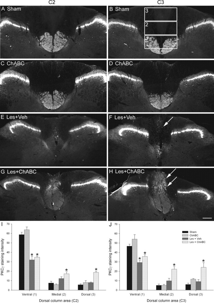
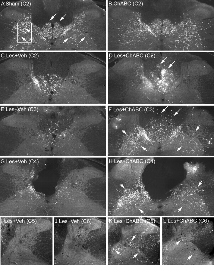
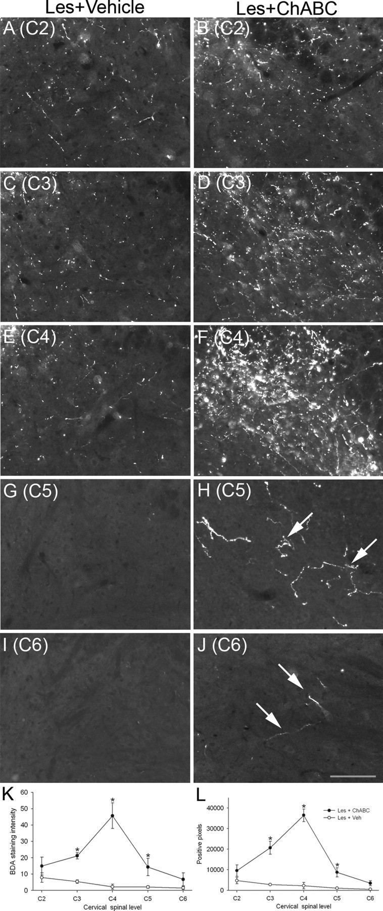
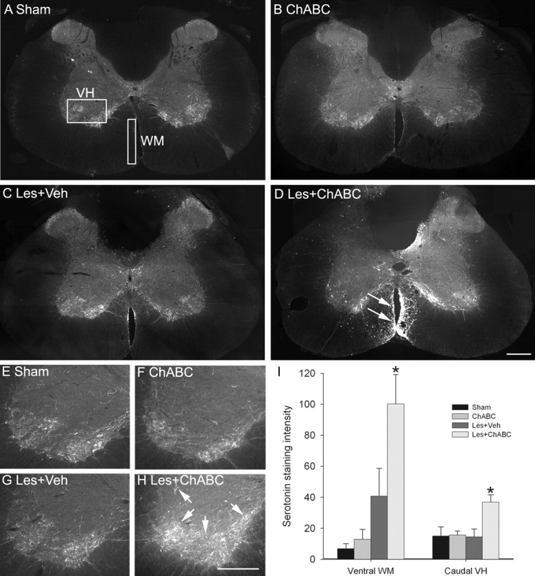
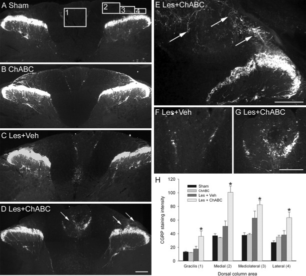
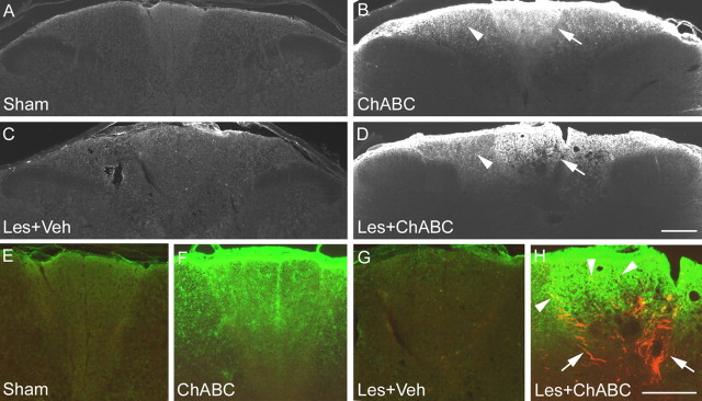
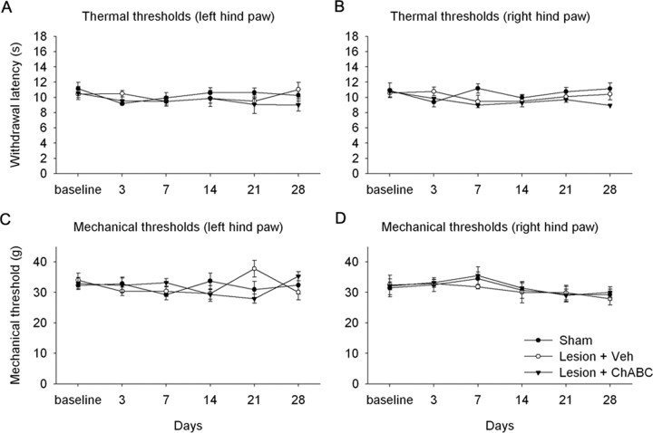
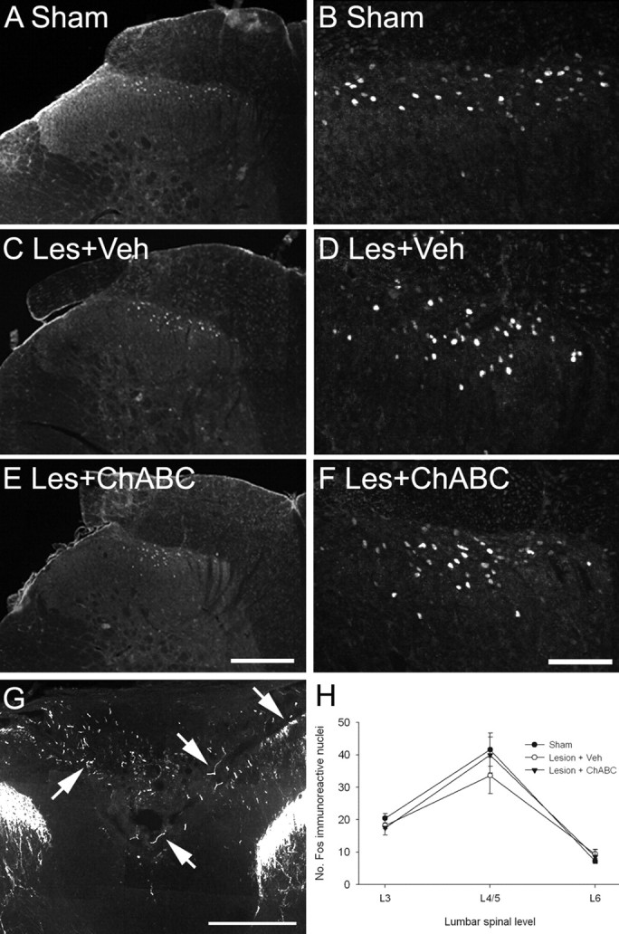
Chondroitin sulfate proteoglycans (CSPGs) are inhibitory extracellular matrix molecules that are upregulated after CNS injury. Degradation of CSPGs using the enzyme chondroitinase ABC (ChABC) can promote functional recovery after spinal cord injury. However, the mechanisms underlying this recovery are not clear. Here we investigated the effects of ChABC treatment on promoting plasticity within the spinal cord. We found robust sprouting of both injured (corticospinal) and intact (serotonergic) descending projections as well as uninjured primary afferents after a cervical dorsal column injury and ChABC treatment. Sprouting fibers were observed in aberrant locations in degenerating white matter proximal to the injury in regions where CSPGs had been degraded. Corticospinal and serotonergic sprouting fibers were also observed in spinal gray matter at and below the level of the lesion, indicating increased innervation in the terminal regions of descending projections important for locomotion. Spinal-injured animals treated with a vehicle solution showed no significant sprouting. Interestingly, ChABC treatment in uninjured animals did not induce sprouting in any system. Thus, both denervation and CSPG degradation were required to promote sprouting within the spinal cord. We also examined potential detrimental effects of ChABC-induced plasticity. However, although primary afferent sprouting was observed after lumbar dorsal column lesions and ChABC treatment, there was no increased connectivity of nociceptive neurons or development of mechanical allodynia or thermal hyperalgesia. Thus, CSPG digestion promotes robust sprouting of spinal projections in degenerating and denervated areas of the spinal cord; compensatory sprouting of descending systems could be a key mechanism underlying functional recovery.


Figures

Figure 1.
Figure 1.
ChABC promotes plasticity of the CST rostral to a C4 dorsal column lesion. A–H, PKCγ immunostaining in transverse sections of the cervical spinal cord at spinal levels C2 (A, C, E, G) and C3 (B, D, F, H) reveals the normal projection of the CST in the ventral part of the dorsal columns in sham (unlesioned) animals (A, B) and ChABC-treated unlesioned animals (C, D). Four weeks after a C4 dorsal column lesion, the CST was mostly absent in the two segments above the injury level after vehicle control treatment (E, F), indicating significant die-back of this tract after injury. CST die-back rostral to injury was also apparent in lesioned animals treated with ChABC; however, in these animals, the CST was observed sprouting into the denervated dorsalmost parts of the dorsal columns, where the ascending projection normally runs (G, H). Arrows indicate the lack of CST growth after lesion and vehicle treatment (F) compared with the robust CST sprouting after lesion and ChABC treatment (H) at the C3 level. Boxed areas in B denote regions selected for quantification. Scale bar, 200 μm. I, J, Quantification in three defined regions at levels C2 (I) and C3 (J) confirmed a significant reduction in PKCγ in the ventral portion of the dorsal columns after injury and de novo expression of PKCγ in the medial and dorsal portions of the dorsal columns after ChABC, but not vehicle, treatment. The asterisks denote a significant difference from sham controls. Error bars indicate SEM. Veh, Vehicle; Les, lesion.
Figure 2.
Figure 2.
ChABC promotes plasticity of CST fibers rostral, within, and caudal to a C4 dorsal column lesion. A, BDA tracing in transverse sections of the cervical spinal cord in sham (uninjured) controls reveals punctate labeling of the CST projection in the ventral portion of the dorsal columns (long arrows) and numerous collaterals innervating spinal gray matter (short arrows). The boxed area denotes the area selected for quantification of CST sprouting in gray matter (see Fig. 3). B, A similar pattern is observed in uninjured animals treated with ChABC. C, E, Serial reconstruction from C2–C6 in lesioned animals treated with vehicle reveals die-back of the CST projection in the dorsal columns as well as decreased innervation of gray matter rostral to the injury at levels C2 and C3. D, F, In contrast, after ChABC treatment, there is reduced CST die-back, numerous intensely labeled bundles of CST axons sprouting dorsally in the dorsal columns (long arrows), and increased CST fibers innervating spinal gray matter (short arrows) rostral to the injury. G, H, At the lesion epicenter (C4), the difference in fiber sprouting within spinal gray matter is compelling with sparse innervation after vehicle treatment (G) and abundant innervation after ChABC treatment, despite a complete absence of the CST (H). I, J, Caudal to the lesion, few, if any, CST fibers were observed in gray matter after vehicle treatment. K, L, However, after ChABC treatment, numerous fibers were observed at level C5 (K) and a small number of fibers were still apparent at C6 (L). Scale bar, 200 μm. Veh, Vehicle; Les, lesion.
Figure 3.
Figure 3.
ChABC promotes plasticity of CST fibers rostral, within, and caudal to a C4 dorsal column lesion. A–J, High-power images of BDA labeling in the spinal gray matter of lesioned animals treated with vehicle (A, C, E, G, I) or ChABC (B, D, F, H, J) showing representative examples of CST fiber sprouting at spinal levels C2–C6. Decreased innervation was apparent rostral to the C4 injury, and no fibers were observed in caudal levels after vehicle treatment; in contrast robust, CST sprouting was apparent rostral to and at the level of the C4 lesion site and regenerating fibers were observed in caudal levels after ChABC treatment. K, L, Quantification of both BDA staining intensity (K) and the number of positive pixels above a threshold (L) revealed a significant increase in CST fibers innervating spinal gray matter after ChABC treatment compared with vehicle treatment, with maximal sprouting occurring at the lesion epicenter (C4) and significant sprouting at both rostral and caudal spinal levels. Asterisks denote a significant difference from sham controls. Scale bar, 200 μm. Les, Lesion; Veh, vehicle. Error bars indicate SEM.
Figure 4.
Figure 4.
ChABC promotes plasticity of serotonergic fibers ventral and caudal to a C4 dorsal column lesion. A, B, Serotonin immunostaining in transverse sections of C4 spinal cord reveals serotonergic immunoreactivity to be expressed predominantly within the superficial dorsal horn, lamina X, and the ventral horn, with a similar pattern of expression observed in sham (unlesioned) animals (A) and ChABC-treated unlesioned animals (B). The boxed areas in A denote regions selected for quantification. C, After dorsal column lesion and vehicle treatment, serotonergic immunoreactivity is similar to that of controls, with a slight increase observed in the ventral white matter adjacent to the ventral sulcus. D, However, after dorsal column lesion and ChABC treatment, there is abundant sprouting of descending serotonergic fibers ventral to the lesion, with numerous intensely stained fibers apparent in ventral white matter (arrows). E–G, Serotonergic innervation caudal to the lesion, within the C6 ventral horn, reveals a similar pattern of innervation in sham (unlesioned) animals (E), ChABC-treated unlesioned animals (F), and lesioned animals treated with vehicle (G). H, In contrast, abundant serotonergic innervation of C6 ventral horn can be observed in lesioned animals treated with ChABC, with dense innervation of serotonergic terminal regions apparent (arrows). I, Quantification confirmed a significant increase in serotonergic immunoreactivity in the ventral white matter at C4 and caudal ventral horn at C6 after ChABC but not vehicle treatment. Asterisks denote a significant difference from sham controls. Error bars indicate SEM. Scale bars, 200 μm. VH, Ventral horn; WM, white matter; Les, lesion; Veh, vehicle.
Figure 5.
Figure 5.
ChABC promotes plasticity of primary afferents caudal to a C4 dorsal column lesion. A, B, CGRP immunostaining in transverse sections of the cervical spinal cord at spinal level C5 reveals the normal pattern of primary afferent terminals in the superficial dorsal horn of sham (unlesioned) animals (A) and ChABC-treated unlesioned animals (B). The boxed areas in A denote regions selected for quantification. C, F, After a C4 dorsal column lesion and vehicle treatment, the pattern of CGRP immunoreactivity at level C5 was similar to that of controls. D, E, G, However, in lesioned animals treated with ChABC, primary afferent sprouting was abundant, particularly in the degenerating dorsal columns, with numerous CGRP-immunoreactive fibers apparent within the gracile fasciculus and in superficial regions of the cuneate fasciculus, where these fibers are not normally present (arrows depict sprouting CGRP-immunoreactive fibers). H, Quantification in four defined regions confirmed a significant increase in CGRP immunoreactivity in the gracile fasciculus and in medial, mediolateral, and lateral areas of the cuneate fasciculus after ChABC but not vehicle treatment. Asterisks denote a significant difference from sham controls. Error bars indicate SEM. Scale bars, 200 μm. Les, Lesion; Veh, vehicle.
Figure 6.
Figure 6.
Sprouting occurs in areas of CSPG digestion after SCI. A, C, In C5 transverse sections, immunostaining for the C-4-S epitope (which is only present after successful digestion of CSPG GAGs) reveals no positive immunoreactivity in sham (uninjured) controls (A) and lesioned animals treated with vehicle (C). B, D, In unlesioned (B) and lesioned (D) animals treated with ChABC, intense C-4-S immunoreactivity is apparent in the dorsal columns, localized in superficial regions of the cuneate fasciculus (arrowheads) and in the gracile fasciculus (arrows), where immunoreactivity was particularly intense (an almost identical pattern was observed in C4 segments; data not shown). E, G, Colocalization of C-4-S (green) and CGRP (red) in high-power images of the graciile fasciculus show no positive staining in sham (uninjured) controls (E) and lesioned animals treated with vehicle (G). F, H, However, although the degree of CSPG digestion in unlesioned (F) and lesioned (H) animals treated with ChABC is comparable, primary afferent sprouting only occurs in the lesioned animals, with numerous CGRP-positive fibers apparent sprouting toward and within areas of CSPG digestion (H, arrows). Scale bars, 200 μm. Les, Lesion; Veh, vehicle.
Figure 7.
Figure 7.
ChABC treatment does not lead to enhanced pain sensitivity after lumbar (L2) dorsal column injury. Group mean latencies for thermal (A, B) or mechanical (C, D) withdrawal thresholds over a 4 week postinjury testing period revealed that there was no development of thermal hyperalgesia or mechanical allodynia after dorsal column lesion and treatment with either vehicle (Veh) or ChABC. Thus, enhanced primary afferent sprouting after injury and ChABC treatment does not result in an increased sensitivity to pain. Error bars indicate SEM.
Figure 8.
Figure 8.
ChABC treatment does not lead to increased connectivity of nociceptive neurons after lumbar (L2) dorsal column injury. Transverse sections through the lumbar spinal cord (level L4/5) show c-fos expression in the dorsal horn 2 h after application of a noxious heat stimulus to the left hindpaw. A, B, In sham controls, noxious heat resulted in the induction of the immediate-early gene c-fos, with c-fos-immunoreactive nuclei apparent in the superficial laminas of the dorsal horn (A; shown in high power in B). C–F, After dorsal column injury and treatment with either vehicle (C, D) or ChABC (E, F), c-fos expression remained unchanged, with a similar pattern of expression to that of controls. G, CGRP immunoreactivity in the lumbar cord of a lesioned animal treated with ChABC reveals that, similar to the observations in the cervical cord, primary afferent sprouting also occurred below the level of a lumbar dorsal column injury. H, Cell counts at lumbar spinal levels L3–L6 confirmed that there were no significant differences in the number of c-fos-immunoreactive nuclei between groups, with c-fos expression maximal at levels L4/5 and fewer c-fos-immunoreactive nuclei apparent in L3 and L6 in all cases. Scale bars: A, C, E, G, 200 μm; B, D, F, 100 μm. Les, Lesion; Veh, vehicle. Error bars indicate SEM.

References

    1. Abbadie C, Skinner K, Mitrovic I, Basbaum AI. Neurons in the dorsal column white matter of the spinal cord: complex neuropil in an unexpected location. Proc Natl Acad Sci USA. 1999;96:260–265. - PMC - PubMed
    1. Asher RA, Morgenstern DA, Fidler PS, Adcock KH, Oohira A, Braistead JE, Levine JM, Margolis RU, Rogers JH, Fawcett JW. Neurocan is upregulated in injured brain and in cytokine-treated astrocytes. J Neurosci. 2000;20:2427–2438. - PMC - PubMed
    1. Asher RA, Morgenstern DA, Shearer MC, Adcock KH, Pesheva P, Fawcett JW. Versican is upregulated in CNS injury and is a product of oligodendrocyte lineage cells. J Neurosci. 2002;22:2225–2236. - PMC - PubMed
    1. Bareyre FM, Kerschensteiner M, Raineteau O, Mettenleiter TC, Weinmann O, Schwab ME. The injured spinal cord spontaneously forms a new intraspinal circuit in adult rats. Nat Neurosci. 2004;7:269–277. - PubMed
    1. Bennett AD, Chastain KM, Hulsebosch CE. Alleviation of mechanical and thermal allodynia by CGRP(8-37) in a rodent model of chronic central pain. Pain. 2000;86:163–175. - PubMed

Publication types

Substances