

Pain processing is faster than tactile processing in the human brain
- PMID: 17050725
- PMCID: PMC6674749
- DOI: 10.1523/JNEUROSCI.2386-06.2006
Pain processing is faster than tactile processing in the human brain
Abstract
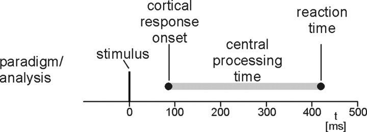
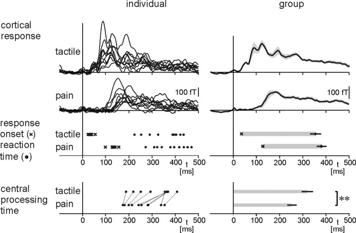
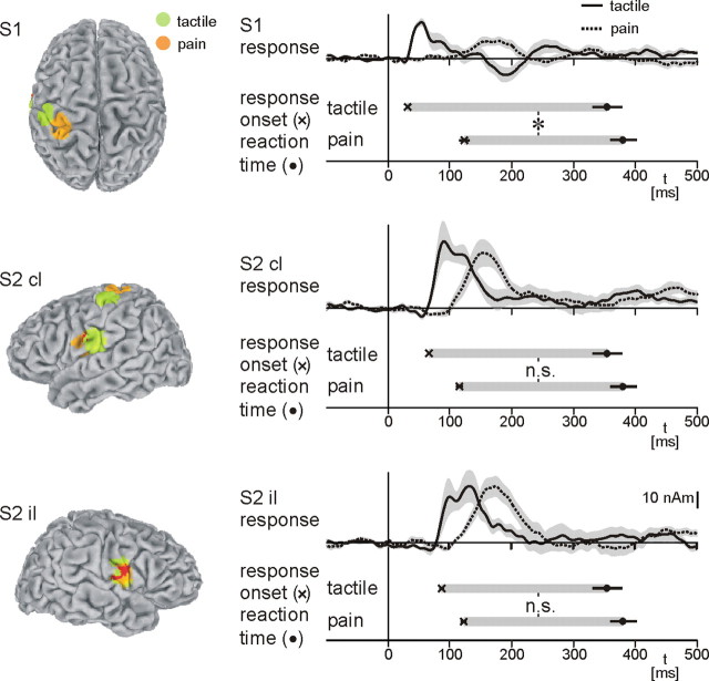
Pain signals threat and drives the individual into a behavioral response that significantly depends on a short stimulus-response latency. Paradoxically, the peripheral and spinal conduction velocities of pain are much slower than of tactile information. However, cerebral processing times and reaction times of touch and pain have not yet been fully assessed. Here we show that reaction times to selective nociceptive cutaneous laser stimuli are substantially faster than expected from the peripheral conduction velocities. Furthermore, by using magnetoencephalography, we found that latencies between earliest stimulus-evoked cortical responses and reaction times are approximately 60 ms shorter for nociceptive than for tactile stimuli. These findings reveal that cerebral processing of pain is substantially faster than processing of tactile information and relatively compensates for the slow peripheral and spinal conduction velocities of pain. Our observation shows how the cerebral organization of pain processing enhances motor responses to potentially harmful stimuli and thereby subserves the particular behavioral demands of pain.
Figures
References
-
- Arendt-Nielsen L, Bjerring P. Reaction times to painless and painful CO2 and argon laser stimulation. Eur J Appl Physiol Occup Physiol. 1988;58:266–273. - PubMed
-
- Binkofski F, Buccino G, Posse S, Seitz RJ, Rizzolatti G, Freund H. A fronto-parietal circuit for object manipulation in man: evidence from an fMRI-study. Eur J Neurosci. 1999;11:3276–3286. - PubMed
-
- Brebner JMT, Welford AT. Introduction: an historical background sketch. In: Welford AT, editor. Reaction times. London: Academic; 1980. pp. 1–23.
-
- Bromm B, Treede RD. Nerve fibre discharges, cerebral potentials and sensations induced by CO2 laser stimulation. Hum Neurobiol. 1984;3:33–40. - PubMed
-
- Bushnell MC, Apkarian AV. Representation of pain in the brain. In: McMahon SB, Koltzenburg M, editors. Wall and Melzack's textbook of pain. Philadelphia: Elsevier; 2006. pp. 107–124.
Publication types
MeSH terms
LinkOut - more resources
Full Text Sources
Other Literature Sources
Medical