. 2006 Nov;188(21):7344-53.
doi: 10.1128/JB.00779-06.

Involvement of nitric oxide in biofilm dispersal of Pseudomonas aeruginosa

Affiliations

Involvement of nitric oxide in biofilm dispersal of Pseudomonas aeruginosa

Nicolas Barraud et al. J Bacteriol. 2006 Nov.

Abstract

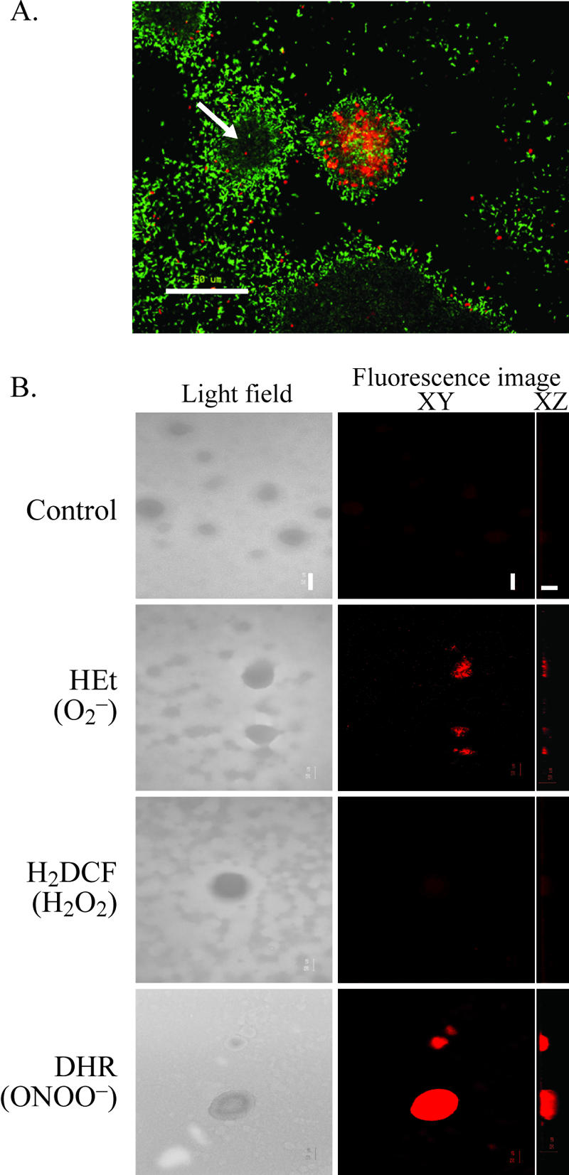
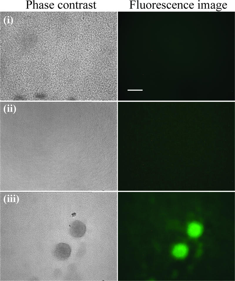
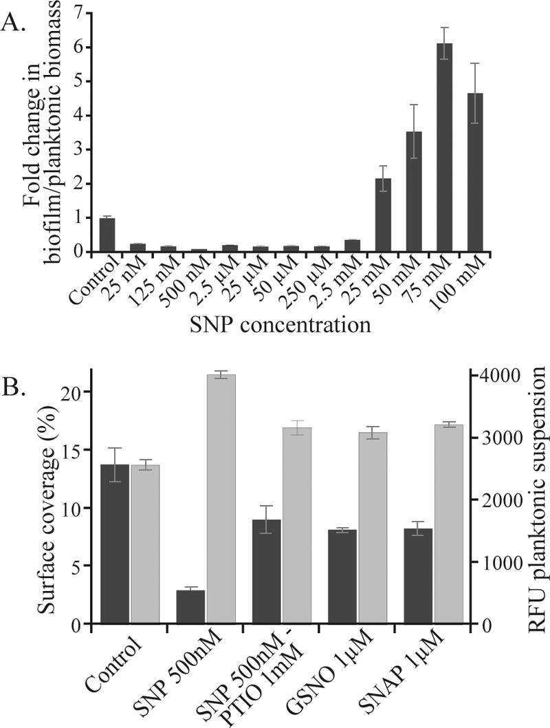
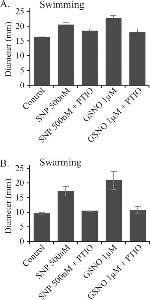
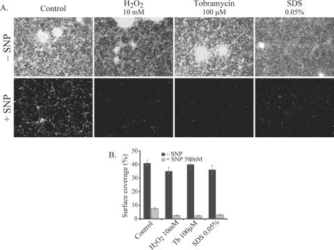
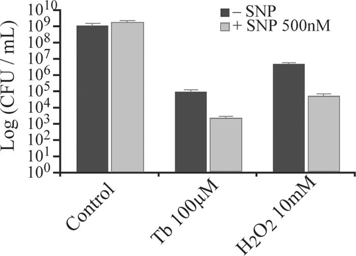
Bacterial biofilms at times undergo regulated and coordinated dispersal events where sessile biofilm cells convert to free-swimming, planktonic bacteria. In the opportunistic pathogen Pseudomonas aeruginosa, we previously observed that dispersal occurs concurrently with three interrelated processes within mature biofilms: (i) production of oxidative or nitrosative stress-inducing molecules inside biofilm structures, (ii) bacteriophage induction, and (iii) cell lysis. Here we examine whether specific reactive oxygen or nitrogen intermediates play a role in cell dispersal from P. aeruginosa biofilms. We demonstrate the involvement of anaerobic respiration processes in P. aeruginosa biofilm dispersal and show that nitric oxide (NO), used widely as a signaling molecule in biological systems, causes dispersal of P. aeruginosa biofilm bacteria. Dispersal was induced with low, sublethal concentrations (25 to 500 nM) of the NO donor sodium nitroprusside (SNP). Moreover, a P. aeruginosa mutant lacking the only enzyme capable of generating metabolic NO through anaerobic respiration (nitrite reductase, DeltanirS) did not disperse, whereas a NO reductase mutant (DeltanorCB) exhibited greatly enhanced dispersal. Strategies to induce biofilm dispersal are of interest due to their potential to prevent biofilms and biofilm-related infections. We observed that exposure to SNP (500 nM) greatly enhanced the efficacy of antimicrobial compounds (tobramycin, hydrogen peroxide, and sodium dodecyl sulfate) in the removal of established P. aeruginosa biofilms from a glass surface. Combined exposure to both NO and antimicrobial agents may therefore offer a novel strategy to control preestablished, persistent P. aeruginosa biofilms and biofilm-related infections.


Figures

FIG. 1.
FIG. 1.
Cell death and dispersal events in P. aeruginosa biofilms correlate with enhanced levels of ONOO inside microcolony structures. (A) Cell death inside biofilm structures occurs simultaneously with biofilm dispersal, as indicated by the formation of hollow biofilm structures (arrow). The image shows a 7-day-old biofilm stained with LIVE/DEAD BacLight stain. Live cells are green, and dead cells are red. (B) Analysis of ROI and RNI in 7-day-old biofilms by use of reactive fluorescent dyes. Images were taken using a confocal microscope; the left panel shows light-field images, and the right panel shows laser scanning fluorescence images showing XY (top) and XZ (side) views. Bacteria in the biofilm showed a low level of autofluorescence, as revealed by the control treatments. Bar, 50 μm.
FIG. 2.
FIG. 2.
Expression of nirS in P. aeruginosa biofilms. Expression of GFP under the control of the nirS (NO2 reductase) promoter was viewed using epifluorescence microscopy; the left panel shows phase-contrast images, and the right panel shows green fluorescence (GFP) images. (i) Aerobically grown planktonic cells (control), (ii) 1-day-old biofilm showing confluent layer of cells on the glass substratum, and (iii) mature biofilms harboring microcolonies are shown. Bar, 50 μm.
FIG. 3.
FIG. 3.
Biofilm development and dispersal of P. aeruginosa wild-type, nitrite reductase-deficient mutant (ΔnirS, unable to produce NO), and NO reductase-deficient mutant (ΔnorCB) strains. (A) Viable cells in the effluent runoff of the wild-type, ΔnirS mutant, and ΔnorCB mutant strain biofilms were enumerated by performing CFU counts. (B) Biofilms were grown in flow cells and stained with LIVE/DEAD staining. Live cells are green, and dead cells are red. Images were collected using a confocal microscope; the upper panel depicts XY (top) views, and the lower panel reveals the XZ (side) axis. After 2 days, all strains show normal biofilm development (i, ii, and iii). After 6 days, the ΔnirS mutant (v) shows a thick biofilm without major dispersal events or cell death, and the ΔnorCB mutant (vi) exhibits extensive detachment and dispersal from the glass surface and numerous hollow voids within the biofilm structure (arrow). Cell death was also greatly enhanced in the ΔnorCB mutant (vi) compared to the wild type (iv). (C) Six-day-old biofilms were stained with the metabolic dye CTC and observed using confocal microscopy. The wild-type strain (vii) and, to a greater extent, the ΔnorCB mutant strain (ix) exhibit high levels of fluorescence in mature microcolonies, whereas ΔnirS mutant biofilm cells (viii) show uniform fluorescence. These images suggest that a subpopulation of cells continue to be active after cell lysis in the microcolonies. Bar, 50 μm.
FIG. 4.
FIG. 4.
NO mediates a transition from biofilm to planktonic mode of growth in P. aeruginosa. (A) PAO1-GFP grown for 24 h in 96-well plates in the presence of SNP. The number of planktonic cells is quantified by fluorescence measurement and biofilm biomass by crystal violet staining. (B) PAO1-GFP grown for 24 h in petri dishes containing microscope slides in the presence of different NO donors (SNP, GSNO, and SNAP) and the scavenger PTIO. Planktonic growth (light-gray bars) was assessed by measuring RFU of the supernatant, and biofilm growth (dark-gray bars) was assessed by measuring the percentage of surface coverage. Error bars indicate standard deviations.
FIG. 5.
FIG. 5.
NO effects on motility behavior in P. aeruginosa. Low concentrations of NO donors (500 nM SNP and 1 μM GSNO) and scavengers (1 mM PTIO) were diluted in motility assay agar plates in triplicate. Migration pattern diameters were measured after 12 to 16 h of swimming (A) and swarming (B). Error bars indicate standard errors.
FIG. 6.
FIG. 6.
NO treatment reverses biofilm formation in P. aeruginosa. Cells remaining on the surface are easily removed by various antimicrobials (tobramycin [Tb], H2O2, and SDS). P. aeruginosa PAO1 was grown in petri dishes containing glass microscope slides. Preestablished biofilms that were grown for 24 h without SNP were allowed to grow for an additional 24 h with (+) or without (−) 500 nM SNP; then, the biofilms on the slides were treated for 30 min with the antimicrobial agents, stained with LIVE/DEAD staining to allow analysis using fluorescence microscopy, and quantified (percent surface coverage) using digital image analysis. (A) The images show microscopic pictures of the biofilms on the glass slides after the combinatorial treatments. (B) The bars show the levels of biofilm biomass after antimicrobial treatment when grown without or with 500 nM SNP, and error bars indicate standard errors.
FIG. 7.
FIG. 7.
NO treatment increases the sensitivity of dispersed P. aeruginosa planktonic cells to antimicrobials (tobramycin [Tb] or H2O2). After 24 h of growth in petri dishes without (−) or with (+) 500 nM SNP, planktonic cells in the supernatant were treated for 2 h with the antimicrobial solutions; then, CFU plate counts were performed to assess the viability of the bacteria. Error bars indicate standard errors.

Comment in

References

    1. Aravind, L., V. Anantharaman, and L. M. Iyer. 2003. Evolutionary connections between bacterial and eukaryotic signaling systems: a genomic perspective. Curr. Opin. Microbiol. 6:490-497. - PubMed
    1. Beckman, J. S., T. W. Beckman, J. Chen, P. A. Marshall, and B. A. Freeman. 1990. Apparent hydroxyl radical production by peroxynitrite: implications for endothelial injury from nitric oxide and superoxide. Proc. Natl. Acad. Sci. USA 87:1620-1624. - PMC - PubMed
    1. Beckman, J. S., and W. H. Koppenol. 1996. Nitric oxide, superoxide, and peroxynitrite: the good, the bad, and ugly. Am. J. Physiol. 271:C1424-C1437. - PubMed
    1. Bindokas, V. P., J. Jordan, C. C. Lee, and R. J. Miller. 1996. Superoxide production in rat hippocampal neurons: selective imaging with hydroethidine. J. Neurosci. 16:1324-1336. - PMC - PubMed
    1. Boyd, A., and A. M. Chakrabarty. 1994. Role of alginate lyase in cell detachment of Pseudomonas aeruginosa. Appl. Environ. Microbiol. 60:2355-2359. - PMC - PubMed

Publication types

MeSH terms