

Death receptor-associated pro-apoptotic signaling in aged skeletal muscle
- PMID: 17051337
- PMCID: PMC5271588
- DOI: 10.1007/s10495-006-0194-6
Death receptor-associated pro-apoptotic signaling in aged skeletal muscle
Abstract
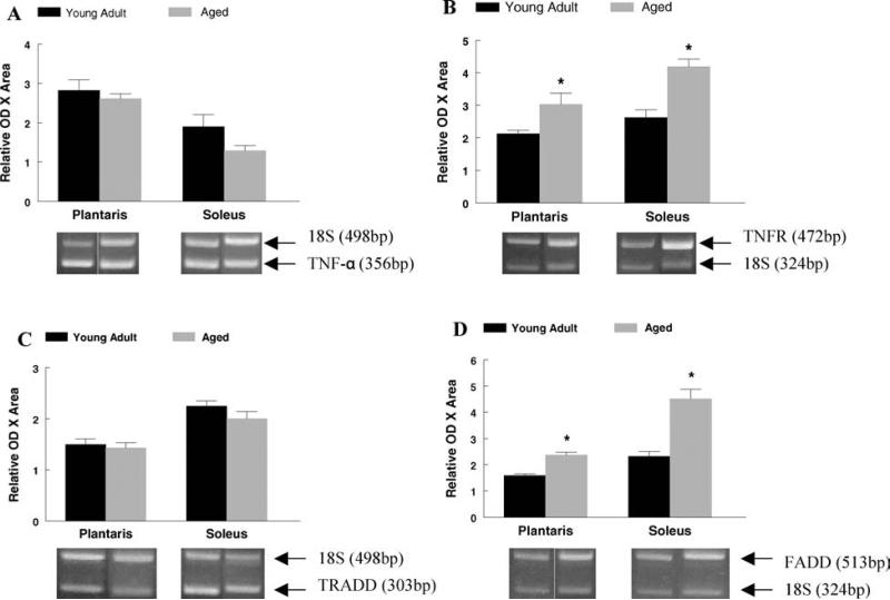
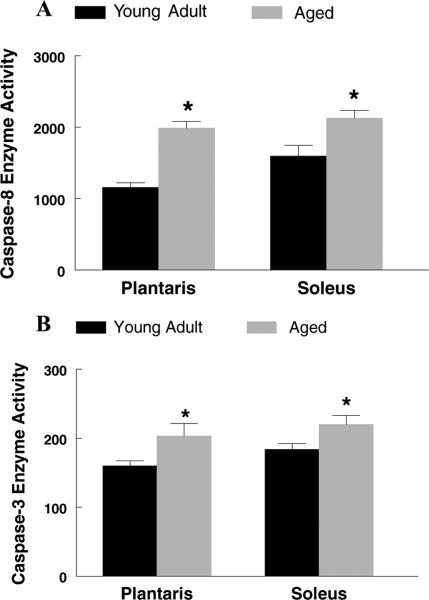
Tumor necrosis factor-alpha (TNF-alpha) is elevated in the serum as a result of aging and it promotes pro-apoptotic signaling upon binding to the type I TNF receptor. It is not known if activation of this apoptotic pathway contributes to the well-documented age-associated decline in muscle mass (i.e. sarcopenia). We tested the hypothesis that skeletal muscles from aged rodents would exhibit elevations in markers involved in the extrinsic apoptotic pathway when compared to muscles from young adult rodents, thereby contributing to an increased incidence of nuclear apoptosis in these muscles. The plantaris (fast) and soleus (slow) muscles were studied in young adult (5-7 mo, n=8) and aged (33 mo, n=8) Fischer(344) x Brown Norway rats. Muscles from aged rats were significantly smaller while exhibiting a greater incidence of apoptosis. Furthermore, muscles from aged rats had higher type I TNF receptor and Fas associated death domain protein (FADD) mRNA, protein contents for FADD, BCL-2 Interacting Domain (Bid), FLICE-inhibitory protein (FLIP), and enzymatic activities of caspase-8 and caspase-3 than muscles from young adult rats. Significant correlations were observed in the plantaris muscle between caspase activity and muscle weight and the apoptotic index, while similar relationships were not found in the soleus. These data demonstrate that pro-apoptotic signaling downstream of the TNF receptor is active in aged muscles. Furthermore, our data extend the previous demonstration that type II fibers are preferentially affected by aging and support the hypothesis that type II fiber containing skeletal muscles may be more susceptible to muscle mass loses via the extrinsic apoptotic pathway.
Figures


Similar articles
-
Changes in IL-15 expression and death-receptor apoptotic signaling in rat gastrocnemius muscle with aging and life-long calorie restriction.Mech Ageing Dev. 2009 Apr;130(4):272-80. doi: 10.1016/j.mad.2008.12.008. Mech Ageing Dev. 2009. PMID: 19396981 Free PMC article.
-
Effects of short-term GH supplementation and treadmill exercise training on physical performance and skeletal muscle apoptosis in old rats.Am J Physiol Regul Integr Comp Physiol. 2008 Feb;294(2):R558-67. doi: 10.1152/ajpregu.00620.2007. Epub 2007 Nov 14. Am J Physiol Regul Integr Comp Physiol. 2008. PMID: 18003794
-
Epigallocatechin-3-gallate improves plantaris muscle recovery after disuse in aged rats.Exp Gerontol. 2014 Feb;50:82-94. doi: 10.1016/j.exger.2013.11.011. Epub 2013 Dec 3. Exp Gerontol. 2014. PMID: 24316035 Free PMC article.
-
The roles of FADD in extrinsic apoptosis and necroptosis.BMB Rep. 2012 Sep;45(9):496-508. doi: 10.5483/bmbrep.2012.45.9.186. BMB Rep. 2012. PMID: 23010170 Review.
-
The role of Ubiquitination in Apoptosis and Necroptosis.Cell Death Differ. 2022 Feb;29(2):272-284. doi: 10.1038/s41418-021-00922-9. Epub 2021 Dec 15. Cell Death Differ. 2022. PMID: 34912054 Free PMC article. Review.
Cited by
-
Detraining Effects of COVID-19 Pandemic on Physical Fitness, Cytokines, C-Reactive Protein and Immunocytes in Men of Various Age Groups.Int J Environ Res Public Health. 2022 Feb 6;19(3):1845. doi: 10.3390/ijerph19031845. Int J Environ Res Public Health. 2022. PMID: 35162870 Free PMC article.
-
TNF-α contributes to sarcopenia through caspase-8/caspase-3/GSDME-mediated pyroptosis.Cell Death Discov. 2023 Feb 24;9(1):76. doi: 10.1038/s41420-023-01365-6. Cell Death Discov. 2023. PMID: 36823174 Free PMC article.
-
The effect of intra-articular injection of different concentrations of ozone on the level of TNF-α, TNF-R1, and TNF-R2 in rats with rheumatoid arthritis.Rheumatol Int. 2013 May;33(5):1223-7. doi: 10.1007/s00296-012-2529-7. Epub 2012 Oct 2. Rheumatol Int. 2013. PMID: 23052485
-
Macrophages and Stem Cells-Two to Tango for Tissue Repair?Biomolecules. 2021 May 6;11(5):697. doi: 10.3390/biom11050697. Biomolecules. 2021. PMID: 34066618 Free PMC article. Review.
-
Pharmacological blockade of TNFα prevents sarcopenia and prolongs survival in aging mice.Aging (Albany NY). 2020 Nov 26;12(23):23497-23508. doi: 10.18632/aging.202200. Epub 2020 Nov 26. Aging (Albany NY). 2020. PMID: 33260150 Free PMC article.
References
-
- Ansved T, Larsson L. Effects of ageing on enzyme-histochemical, morphometrical and contractile properties of the soleus muscle in the rat. J Neurol Sci. 1989;93:105–124. - PubMed
-
- Larsson L, Grimby G, Karlsson J. Muscle strength and speed of movement in relation to age and muscle morphology. J Appl Physiol. 1979;46:451–456. - PubMed
-
- Larsson L, Sjodin B, Karlsson J. Histochemical and biochemical changes in human skeletal muscle with age in sedentary males, age 22–65 years. Acta Physiol Scand. 1978;103:31–39. - PubMed
-
- Tenover JL. Testosterone and the aging male. J Androl. 1997;18:103–106. - PubMed
-
- Tenover JS, Matsumoto AM, Plymate SR, Bremner WJ. The effects of aging in normal men on bioavailable testosterone and luteinizing hormone secretion: response to clomiphene citrate. J Clin Endocrinol Metab. 1987;65:1118–1126. - PubMed
Publication types
MeSH terms
Substances
Grants and funding
LinkOut - more resources
Full Text Sources
Medical
Research Materials
Miscellaneous
