. 2007 Jan;120(1):73-82.
doi: 10.1111/j.1365-2567.2006.02479.x. Epub 2006 Oct 18.

Natural killer cell and hepatic cell interaction via NKG2A leads to dendritic cell-mediated induction of CD4 CD25 T cells with PD-1-dependent regulatory activities

Affiliations

Natural killer cell and hepatic cell interaction via NKG2A leads to dendritic cell-mediated induction of CD4 CD25 T cells with PD-1-dependent regulatory activities

Masahisa Jinushi et al. Immunology. 2007 Jan.

Abstract

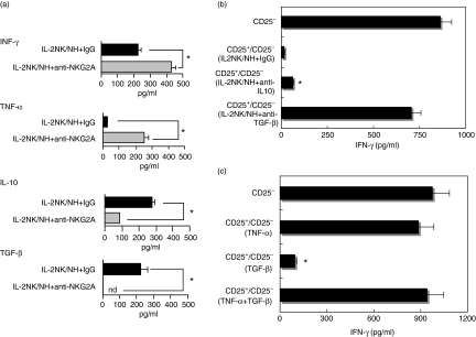
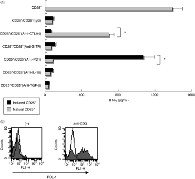
Natural killer (NK) cells have the ability to control dendritic cell (DC)-mediated T cell responses. However, the precise mechanisms by which NK receptor-mediated regulation of NK cells determines the magnitude and direction of DC-mediated T cell responses remain unclear. In the present study, we applied an in vitro co-culture system to examine the impact of NK cells cultured with hepatic cells on DC induction of regulatory T cells. We found that interaction of NK cells and non-transformed hepatocytes (which express HLA-E) via the NKG2A inhibitory receptor resulted in priming of DCs to induce CD4(+) CD25(+) T cells with regulatory properties. NKG2A triggering led to characteristic changes of the cytokine milieu of co-cultured cells; an increase in the transforming growth factor (TGF)-beta involved in the generation of this specific type of DC, and a decrease in the tumour necrosis factor-alpha capable of antagonizing the effect of TGF-beta. The regulatory cells induced by NK cell-primed DCs exert their suppressive actions through a negative costimulator programmed death-1 (PD-1) mediated pathway, which differs from freshly isolated CD4(+) CD25(+) T cells. These findings provide new insight into the role of NK receptor signals in the DC-mediated induction of regulatory T cells.


Figures

Figure 1
Figure 1
Human non-transformed hepatocyte (NH) modulation of activated natural killer (NK) cells endows dendritic cells (DCs) with the ability to induce CD4+ CD25+ regulatory T cells. (a) Freshly isolated CD4+ CD25+ T cells were cultured in the presence of plate-bound anti-CD3 antibody (Ab) for 24 hr, and then subjected to flow cytometry to examine their expression of cytotoxic T lymphocyte antigen-4 (CTLA-4), glucocorticoid-induced TNF receptor (GITR) and programmed death-1 (PD-1) (closed histograms). Open histograms represent the staining of control Ab. Numbers on the upper right indicate the mean fluorescence intensity (MFI) of each type of stained cells. (b) NK cells were preactivated with 50 ng/ml interleukin (IL)-2, and co-cultured in the absence (IL-2 NK) or presence (IL-2 NK/NH) of NHs at a ratio of 1 : 1 for 24 hr. DCs (1 × 10⁵) were stimulated for 24 hr with the supernatant obtained from the co-cultured medium. After washing three times, DCs were cultured with allogeneic CD4+ T cells for 48 hr. CD4+ CD25+ fractions were isolated from the DC/CD4+ T co-culture and subjected to flow cytometry for expression of CTLA-4, GITR or PD-1 (closed histograms). Open histograms show isotype control staining. Numbers on the upper right indicate the MFI of each type of stained cell. (c) All experiments in (a) and (b) were performed three times and the composite results with statistical analysis are shown as the MFI of the staining cells. *P < 0·05 vs. responses of IL-2 NK/NH group. The experiment was performed with a different set of donors and similar results were obtained. (d) PD-1 expression on CD4+ CD25+ T cells stimulated with allogeneic DCs from three different donors, shown as the MFI.(e)CD4+ CD25+ T cells were prepared as described above. The mRNA expression of Foxp3 and GAPDH (glyceraldehyde 3-phosphate dehydrogenase) was examined by reverse transcription-polymerase chain reaction (RT-PCR). (f) CD4+ CD25+ fractions were isolated from DC/CD4+ T cell co-cultures. Different numbers of these CD4+ CD25+ T cells were co-cultured with freshly isolated autologous CD4+ CD25 T cells (1 × 10⁵/well) in the presence of plate-bound anti-CD3 Ab (CD4+ CD25+/CD4+ CD25). The anti-CD3 Ab-activated CD4+ CD25 T cells alone were used as a positive control (CD4+ CD25). IFN-γ was measured for each supernatant obtained after 48 hr of co-culture by enzyme-linked immunosorbent assay. *P < 0·05.
Figure 2
Figure 2
NKG2A signals of natural killer (NK) cells are required for the dendritic cell (DC) induction of CD4+ CD25+ T cells with the regulatory phenotype. (a) Surface expression of the ligands of NKG2A (HLA-E) as well as NKG2D (MIC, ULBP1 and ULBP2) in human non-transformed hepatocytes (NHs) were assessed by flow cytometry (closed histograms). Open histograms show isotype control staining. (b, c) Interleukin (IL)-2-preactivated NK cells were co-cultured with NHs in the presence of 30 µg/ml of anti-NKG2A neutralizing antibody (Ab) (anti-NKG2A) or control IgG. DCs (1 × 10⁵) were then stimulated with the supernatant obtained from the co-cultured medium for 24 hr. After washing three times, DCs were cultured with allogeneic CD4+ T cells for 48 hr. CD4+ CD25+ cells isolated from the co-culture were subjected to FCM for their surface expression of cytotoxic T lymphocyte antigen-4 (CTLA-4), glucocorticoid-induced TNF receptor (GITR) and programmed death-1 (PD-1) (closed histograms). Open histograms show isotype control staining. Numbers on the upper right indicate the mean fluorescence intensity (MFI) of each type of stained cell. All experiments were performed three times. Representative data (b) and composite results with statistical analysis (c) are shown as the MFI of the staining cells. *P < 0·05 vs. responses of IgG group. The experiment was performed in different set of donors and similar results were obtained. (d) The inhibitory effect of anti-NKG2A Ab on PD-1 expression of CD4+ CD25+ T cells stimulated with allogeneic DCs from three different donors. Data are shown as MFI.(e)CD4+ CD25+ T cells were stimulated and purified as described above. The mRNA expression of Foxp3 and GAPDH (glyceraldehyde 3-phosphate dehydrogenase) was examined by reverse transcription-polymerase chain reaction (RT-PCR. (f) CD4+ CD25+ T cells (1 × 10⁵/well) isolated from DC and CD4+ T cell co-cultures were cultured with freshly isolated autologous CD4+ CD25 T cells at a ratio of 1 : 1 in the presence of plate-bound anti-CD3 Ab (CD25+/CD25). The anti-CD3 Ab-activated CD4+ CD25 T cells alone were used as a positive control (CD25). Interferon (IFN)-γ was measured for each supernatant obtained after 48 hr of co-culture by enzyme-linked immunosorbent assay (ELISA). *P < 0·05. All experiments were performed three times; representative results are shown.
Figure 3
Figure 3
Change of cytokine production pattern of natural killer (NK) cells through NKG2A signals is responsible for the dendritic cell (DC) induction of CD4+ CD25+ Treg cells. (a) NK cells prestimulated with interleukin (IL)-2 were cultured with human non-transformed hepatocytes (NHs) in the presence of masking antibodies (Abs) of NKG2A (IL-2 NK/NH + anti-NKG2A) or isotype control IgG (IL-2 NK/NH + IgG) for 24 hr. *P < 0·05. (b) IL-2 activated NK cells were co-cultured with NHs (IL-2 NK/NH). DCs (1 × 10⁵) were stimulated with the culture supernatant in the presence of anti-IL-10, anti-transforming growth factor (TGF)-β neutralizing Ab or control IgG for 24 hr. DCs were washed thoroughly and co-cultured with allogeneic CD4+ T cells for 48 hr. Next, the isolated CD4+ CD25+ T cells (1 × 10⁵/well) were co-cultured with autologous CD4+ CD25 T cells in the presence of plate-bound anti-CD3 Ab at a ratio of 1 : 1. Interferon (IFN)-γ production from the culture supernatant was examined by enzyme-linked immunosorbent assay. *P < 0·05 vs. responses of anti-CD3 Ab-stimulated CD4+ CD25 T cells. (c) DCs (1 × 10⁵) were stimulated with 50 ng/ml TNF-α, 100 ng/ml TGF-β or both for 24 hr. After thorough washing, they were co-cultured with allogeneic CD4+ T cells for 48 hr. CD4+ CD25+ T cells (1 × 10⁵/well) were isolated from the DC and CD4+ co-cultures and cultured with freshly isolated autologous CD4+ CD25 T cells at a ratio of 1 : 1 in the presence of plate-bound anti-CD3 Ab. IFN-γ production was examined as described above. *P < 0·05 vs. responses of anti-CD3 Ab-stimulated CD4+ CD25 T cells.
Figure 4
Figure 4
CD4+ CD25+ Treg cells induced by interleukin (IL)-2 natural killer (NK)/human non-transformed hepatocytes (NH)-treated dendritic cell (DC) suppressed T cell activation through programmed death-1 (PD-1)/programmed death ligand-1 (PDL-1) interactions. (a) DCs (1 × 10⁵) were stimulated with the IL-2 NK/NH supernatant for 24 hr, and then cultured with allogeneic CD4+ T cells for 48 hr. CD4+ CD25+ fractions were isolated from the DC/CD4+ T cell mixtures. Freshly isolated CD4+ CD25+ T cells (natural CD25+) or CD4+ CD25+ T cells induced by NK/NH-primed DCs (induced CD25+) were co-cultured with freshly isolated autologous CD4+ CD25 T cells at a ratio of 1 : 1 upon stimulation of plate-bound anti-CD3 antibody (Ab). Anti-CTLA-4 (cytotoxic T lymphocyte antigen-4) Ab, anti-GITR (glucocorticoid-induced TNF receptor) Ab, anti-PD-1 Ab, anti-IL-10 Ab, anti-TGF-β Ab or isotype control IgG (20 µg/ml for each) were incubated during CD4+ CD25+/CD4+ CD25 T cell co-cultures. Interferon (IFN)-γ was measured for each supernatant obtained after 72 hr of co-culture by enzyme-linked immunosorbent assay. *P < 0·05 vs. responses of anti-CD3 Ab-stimulated CD4+ CD25 T cells. (b) Freshly isolated CD4+ CD25 T cells were incubated with (anti-CD3) or without (–) plate-bound anti-CD3 Ab for 24 hr. PDL-1 expression was assessed by flow cytometry (closed histograms). Open histograms show isotype control staining.

Similar articles

Cited by

References

    1. Shevach EM. CD4+ CD25+ suppressor T cells: more questions than answers. Nat Rev Immunol. 2002;2:389–400. - PubMed
    1. Jonuleit H, Schmitt E, Stassen M, Tuettenberg A, Knop J, Enk AH. Identification of functional characterization of human CD4+ CD25+ T cells with regulatory properties isolated from peripheral blood. J Exp Med. 2001;193:1285–94. - PMC - PubMed
    1. Dieckmann D, Plottner H, Berchtold S, Berger T, Schuler G. Ex vivo isolation and characterization of CD4+ CD25+ T cells with regulatory properties from human blood. J Exp Med. 2001;193:1303–10. - PMC - PubMed
    1. Belkald Y, Piccirillo CA, Mendez S, Shevach EM, Sacks DL. CD4+ CD25+ regulatory T cells control Leishmania major persistence and immunity. Nature. 2002;420:502–7. - PubMed
    1. Wang HY, Lee DA, Peng G, Guo Z, Li Y, Kiniwa Y, Shevach EM, Wang RF. Tumour-specific human CD4+ regulatory T cells and their ligands: implication for immunotherapy. Immunity. 2004;20:107–18. - PubMed

Publication types

MeSH terms