. 2006 Oct 20;24(2):233-43.
doi: 10.1016/j.molcel.2006.09.020.

Two functional modes of a nuclear receptor-recruited arginine methyltransferase in transcriptional activation

Affiliations

Two functional modes of a nuclear receptor-recruited arginine methyltransferase in transcriptional activation

María J Barrero et al. Mol Cell. .

Abstract

Nuclear receptors, like other transcriptional activators, switch on gene transcription by recruiting a complex network of coregulatory proteins. Here, we have identified the arginine methyltransferase PRMT1 as a coactivator for HNF4, an orphan nuclear receptor that regulates the expression of genes involved in diverse metabolic pathways. Remarkably, PRMT1, whose methylation activity on histone H4 strongly correlates with induction of HNF4 target genes in differentiating enterocytes, regulates HNF4 activity through a bipartite mechanism. First, PRMT1 binds and methylates the HNF4 DNA-binding domain (DBD), thereby enhancing the affinity of HNF4 for its binding site. Second, PRMT1 is recruited to the HNF4 ligand-binding domain (LBD) through a mechanism that involves the p160 family of coactivators and methylates histone H4 at arginine 3. This, together with recruitment of the histone acetyltransferase p300, leads to nucleosomal alterations and subsequent RNA polymerase II preinitiation complex formation.


Figures
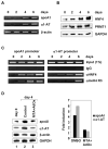
Figure 1
Figure 1. Histone H4 R3 methylation at HNF4 target promoters correlates with HNF4 occupancy during differentiation of CaCo-2 cells
A. mRNA levels of apoAI, α1-AT and β-actin were determined by RT-PCR at the indicated days after cells reached confluence. B. Western blot showing protein levels of HNF4, PRMT1 and GAPDH during differentiation. C. ChIP assays on the α1-AT and apoAI promoters were performed during CaCo-2 differentiation using specific antibodies against HNF4 (αHNF4), histone H4 methylated at R3 (αmeH4-R3), or unrelated IgGs (IgG). D. Effect of MTA and AdOx on the indicated genes mRNA levels during differentiation. Radiolabeled RT-PCR products (left panel) were quantified and apoAI and α1-AT mRNA levels were normalized to β-actin and plotted (right panel) as fold induction (day 4 vs. day 0).
Figure 2
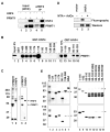
Figure 2. HNF4 interacts directly with PRMT1 through its DNA binding domain and methylates HNF4 at R91
A. The association of coexpressed HNF4 and PRMT1 in 293T cells was analyzed following immunoprecipitation with antibodies against HNF4. B. In vitro interaction of HNF4 and PRMT1 using 2 μg of GST proteins coupled to glutathione-sepharose beads, 0.5 μg of purified His-tagged PRMT1 and 180 mM KCl in the binding and washing buffer. C. In vivo methylation of FLAG-tagged HNF4 or PRMT1 overexpressed in 293T cells and purified using anti-FLAG M2 antibodies. Left and right panels show Coomassie staining and fluorography respectively. Asterisks indicate methylated proteins that copurify with PRMT1. D. In vivo methylation of FLAG-tagged HNF4 in the absence or presence of MTA and AdOx. Methylated HNF4 was detected by fluorography (upper panel) and total HNF4 was detected by western blot (lower panel). E. In vitro methylation assays using 250 ng of His-tagged PRMT1 and 2 μg of the indicated GST proteins. Upper and lower panels show Coomassie and fluorography respectively. Arrows indicate PRMT1.
Figure 3
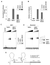
Figure 3. HNF4 methylation increases its binding to DNA
A. FLAG-tagged HNF4 or HNF4 R91W expression plasmids were cotransfected into 293T cells together with ABC.Luc. The plot represents the mean with standard deviation (shown by the error bars) of normalized luciferase activity from four independent experiments. Levels of HNF4 were detected by western blot. B. ChIP assays in 293T cells cotransfected with FLAG-tagged HNF4 or HNF4 R91W and the reporter plasmid Ax2.Luc were performed using HNF4 antibodies. Recruitment relative to control IgGs is represented. Levels of FLAG-tagged HNF4 immunoprecipitated from the crosslinked sample were quantified by dot blot using HNF4 antibodies. C. FLAG-tagged HNF4 or HNF4 R91W overexpressed in 293T were purified using M2 agarose and eluted using FLAG peptide; 5 μl of each was analyzed by western blot with anti-HNF4 antibodies (right panel) and visually estimated to be in the subnanogram range. The purified proteins were tested for their ability to shift an apoAI site A (−214/−192) probe. Lane 1 contains free probe; lane 2 contains immunoprecipitated material from non-transfected cells (control); lanes 3 to 6 and lanes 7 to 10 contain increasing amounts (0.5, 1, 2, 4 μl) of wild type FLAG-tagged HNF4 or HNF4 R91W, respectively. D. His-tagged recombinant proteins were expressed and purified from bacteria, resolved by SDS-PAGE and quantified by Coomassie staining (right panel). Gel shift experiment was performed as in C. Lane 11 contains free probe, lanes 12 to 15 and lanes 16 to 19 contain increasing amounts (6.25, 12.5, 25 and 50 ng) of His-tagged HNF4 or HNF4 R91W, respectively. E. Map of HNF4 DBD showing the residues modified by PRMT1, p300/CBP and PKA. [Adapted from (Viollet et al., 1997)].
Figure 4
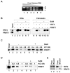
Figure 4. In vitro HNF4 methylation increases its DNA binding activity on chromatin
A. Partial micrococcal nuclease digestion of chrgomatin reconstituted from a biotinylated template containing four HNF4 binding sites. The core histones to DNA ratio varies: 1.1:1 (lane 1); 1:1 (lane 2); 0.75:1 (lane 3); 0.6:1 (lane 4); 0.55:1 (lane 5) and 0.5:1 (lane 6). “a” corresponds to a 450 bp undigested DNA; “b” corresponds to a 300 bp dinucleosome band; “c” corresponds to a 170 bp mononucleosome band. B. Binding of HNF4 and PRMT1 to an immobilized template either reconstituted into chromatin (lanes 8–13) or naked (lanes 1–7), in the absence or presence of SAM, as indicated. C and D. For GST pull-downs, 50 ng of GST-HNF4 (1-175) in C or GST-H4 in D, were incubated with 1 μg of His-tagged PRMT1 for 2 hr at 37ºC in the absence or presence of SAM. Complexes were then pulled down and washed with buffer containing the indicated KCl concentrations. E. HNF4 was incubated with the immobilized DNA template, washed to eliminate unbound HNF4, quantified and subjected to in vitro methylation (lanes 2 and 4) in parallel with an equivalent amount (2 μg) of free HNF4 (lanes 1 and 3). Reactions included 200 ng of His-tagged PRMT1. Coomassie (lanes 1 and 2) and fluorography (lanes 3 and 4) are shown.
Figure 5
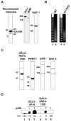
Figure 5. In vitro transcription system for chromatin templates
A. Coomassie staining of the purified proteins used for chromatin reconstitution. B. Partial microccocal nuclease digestion of chromatin reconstituted with wild type (WT, lane 2) or mutant (H4 R3Q, lane 4) core histones with the histone to DNA ratio of 1.1:1. Lanes 1 and 3 show a 123 bp ladder. C. Purified factors used for in vitro transcription. Asterisks indicate non-specific proteins. D. In vitro transcription reactions contained 50 ng of chromatinized template, 10 ng of purified GAL4-VP16, 100 ng of GAL4-HNF4-LBD and 20 ng of p300, as indicated. Transcription data were quantified by phosphorimager and are expressed as relative transcription (Txn), using the lowest detectable signal as reference. ND, not determined.
Figure 6
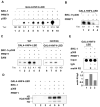
Figure 6. PRMT1 displays synergistic effects with SRC-1 and p300 on HNF4-dependent in vitro transcription of chromatin templates
A. In vitro transcription reactions contained 50 ng of chromatin reconstituted template, 100 ng of GAL4-HNF4-LBD, 50 ng of PRMT1, 20 ng of SRC-1 and 20 ng of p300, as indicated. B. In vitro transcription reactions contained 50 ng of naked template, 100 ng of GAL4-HNF4-LBD, 20 ng of PRMT1, 5 ng of SRC-1 and 5 ng of p300, as indicated. C. In vitro transcription as in B but using 50 ng of template reconstituted into chromatin using wild type recombinant histones or mutant H4 R3Q histones. Reactions were carried out in parallel but different exposure times at comparable baseline intensities are shown. D. Histone acetyltransferase assay on chromatin templates. Reactions contain 100 ng of GAL4-HNF4-LBD, 100 ng of PRMT1, 60 ng of SRC-1 and 60 ng of p300, as indicated. E. In vitro transcription was performed as in C using wild type recombinant histones for chromatin reconstitution and the indicated combination of factors. After transcription, chromatin was immunoprecipitated using antibodies against histone H4 methylated at R3 (meH4 R3), or unrelated IgGs (IgG), dot-blotted and probed with a promoter probe.
Figure 7
Figure 7. HNF4, PRMT1 and SRC-1 form a ternary complex
A. GST pull down using 10 μg of GST proteins coupled to glutathione-sepharose beads, 100 ng of SRC-1 and 150 mM KCl binding and washing buffer. B. GST pull down using 10 μg of GST proteins coupled to glutathione-sepharose beads, 300 ng of SRC-1, 1 μg of PRMT1 and 100 mM KCl in the binding and washing buffer. Inputs (5% for SRC1 and 1% for PRMT1) are also shown. C. Model for PRMT1-regulated HNF4 function. PRMT1 binds directly to the HNF4 DBD and methylates R91, increasing its DNA binding. Once HNF4 is bound to DNA, PRMT1 is recruited by the LBD through SRC-1 and methylates histone H4 at R3. This, together with recruitment of other coactivators like p300, facilitates the PIC formation and the transcription of HNF4 target genes.

References

    1. An W, Kim J, Roeder RG. Ordered cooperative functions of PRMT1, p300, and CARM1 in transcriptional activation by p53. Cell. 2004;117:735–748. - PubMed
    1. An W, Roeder RG. Reconstitution and transcriptional analysis of chromatin in vitro. Methods Enzymol. 2004;377:460–474. - PubMed
    1. Bedford MT, Richard S. Arginine methylation an emerging regulator of protein function. Mol Cell. 2005;18:263–272. - PubMed
    1. Chen D, Huang SM, Stallcup MR. Synergistic, p160 coactivator-dependent enhancement of estrogen receptor function by CARM1 and p300. J Biol Chem. 2000;275:40810–40816. - PubMed
    1. Chen WS, Manova K, Weinstein DC, Duncan SA, Plump AS, Prezioso VR, Bachvarova RF, Darnell JE., Jr Disruption of the HNF-4 gene, expressed in visceral endoderm, leads to cell death in embryonic ectoderm and impaired gastrulation of mouse embryos. Genes Dev. 1994;8:2466–2477. - PubMed

Publication types

MeSH terms