. 2007 Mar 1;109(5):1801-9.
doi: 10.1182/blood-2006-08-043471. Epub 2006 Oct 19.

Redefining endothelial progenitor cells via clonal analysis and hematopoietic stem/progenitor cell principals

Affiliations

Redefining endothelial progenitor cells via clonal analysis and hematopoietic stem/progenitor cell principals

Mervin C Yoder et al. Blood. .

Abstract

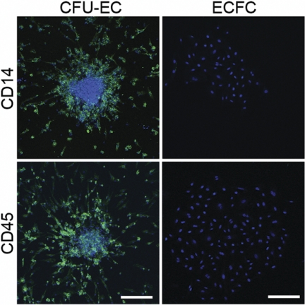
The limited vessel-forming capacity of infused endothelial progenitor cells (EPCs) into patients with cardiovascular dysfunction may be related to a misunderstanding of the biologic potential of the cells. EPCs are generally identified by cell surface antigen expression or counting in a commercially available kit that identifies "endothelial cell colony-forming units" (CFU-ECs). However, the origin, proliferative potential, and differentiation capacity of CFU-ECs is controversial. In contrast, other EPCs with blood vessel-forming ability, termed endothelial colony-forming cells (ECFCs), have been isolated from human peripheral blood. We compared the function of CFU-ECs and ECFCs and determined that CFU-ECs are derived from the hematopoietic system using progenitor assays, and analysis of donor cells from polycythemia vera patients harboring a Janus kinase 2 V617F mutation in hematopoietic stem cell clones. Further, CFU-ECs possess myeloid progenitor cell activity, differentiate into phagocytic macrophages, and fail to form perfused vessels in vivo. In contrast, ECFCs are clonally distinct from CFU-ECs, display robust proliferative potential, and form perfused vessels in vivo. Thus, these studies establish that CFU-ECs are not EPCs and the role of these cells in angiogenesis must be re-examined prior to further clinical trials, whereas ECFCs may serve as a potential therapy for vascular regeneration.


Figures

Figure 1
Figure 1
Culture of EPCs from human peripheral blood. (A) Two methods for isolating and culturing EPCs from human peripheral blood. Yellow cells represent nonadherent cells and red cells represent adherent cells. FN indicates fibronectin. (B) Representative phase-contrast photomicrograph of a CFU-EC colony (day 5) cultured from adult peripheral blood MNCs by method A. Similar colonies were observed from 29 other adult peripheral and 10 cord blood donors. Scale bar represents 500 μm. (C) Representative phase-contrast photo-micrograph of an ECFC colony (day 19) cultured from adult peripheral blood MNCs by method B. Similar colonies were observed from 29 other adult peripheral and 10 cord blood donors. Arrows indicate colony boundary and scale bar represents 500 μm.
Figure 2
Figure 2
CFU-EC and ECFC colonies generated from discarded cells from methods A and B. (A) Representative photomicrograph phase-contrast of an ECFC colony, which arose when the adherent cells discarded from method A were cultured in EGM-2 medium (method B). Similar colonies were observed from 4 other adult peripheral and 5 cord blood donors. Arrows indicate colony boundary and scale bar represents 500 μm. (B) Representative phase-contrast photomicrograph of a CFU-EC colony, which arose when the nonadherent cells discarded from method B were cultured by method A. Similar colonies were observed from 4 other adult peripheral and 5 cord blood donors. Scale bar represents 500 μm.
Figure 3
Figure 3
Expression of endothelial cell antigens by CFU-ECs and ECFCs. (A) Immunophenotyping of CFU-EC and ECFC colonies by confocal microscopy. CFU-ECs and ECFCs express CD31, CD105, CD144, CD146, KDR, VWF, and UEA-I. Shown are isotype and antigen staining (green) representing 5 independent experiments using cells from different donors. Nuclei are stained with Hoechst 33342 (blue) and scale bar represents 200 μm. (B) CFU-ECs and ECFCs incorporate Ac-LDL. Shown is a confocal photomicrograph of cells that have taken up Ac-LDL (green) following a 4-hour incubation, representing 5 independent experiments using cells from different donors. Nuclei are stained with Hoechst 33342 (blue) and scale bar represents 200 μm.
Figure 4
Figure 4
Expression of hematopoietic-specific cell surface antigens by CFU-ECs and ECFCs. Immunophenotyping of CFU-EC and ECFC colonies by confocal microscopy. CFU-ECs but not ECFCs express CD14 and CD45 (green). Photomicrographs are representative of 5 independent experiments using cells from different donors. Nuclei are stained with Hoechst 33342 (blue) and scale bar represents 200 μm.
Figure 5
Figure 5
Monocyte/macrophage function in CFU-ECs. (A) Detection of cell surface expression of CD115 on CFU-EC and ECFC colonies by immunofluorescent staining. CFU-ECs express CD115 (green). Confocal photomicrographs are representative of 5 independent experiments using cells from different donors. Nuclei are stained with Hoechst 33342 (blue) and scale bar represents 200 μm. (B) Representative phase-contrast photomicrograph of CFU-EC colonies exposed to α-naphthyl acetate esterase with and without NaF inhibition. Similar nonspecific esterase activity was seen in CFU-EC colonies from 4 other donors. Scale bar represents 500 μm. (C) RT-PCR analysis of whole peripheral blood MNCs (M), CFU-ECs (C), and ECFCs (E) for gene expression of CD14, CD45, CD115, and β-actin. Left 3 lanes show reactions absent for reverse transcriptase. Results are representative of 5 independent experiments using cells from different donors. (D) Percentage of cells derived from CFU-EC or ECFC colonies that phagocytose E coli. Results represent the mean percentage of cells that phagocytose E coli ± SEM of 5 independent experiments. J774 murine monocytes were used as a positive control. *P < .001 by Student paired t test. (E) CFU-EC–derived cells demonstrate the ability to phagocytose E coli. Representative confocal photomicrographs of J774 murine monocytes, and CFU-EC– and ECFC-derived cells exposed to fluorescein-labeled E coli. Similar results were seen in 4 other experiments with cells derived from different donors. Nuclei are stained with Hoechst 33342 (blue) and scale bar represents 50 μm.
Figure 6
Figure 6
Secondary colony formation. (A) Representative phase-contrast photomicrographs of the cell progeny or secondary colonies (right) formed 7 days after primary CFU-EC and ECFC colonies (left) were plated at low cell density. Similar results were seen in 99 other CFU-EC colonies and 59 other ECFC colonies. Scale bar represents 500 μm. (B) Representative phase-contrast photomicrograph (original magnification, × 10) of secondary CFU-GM colonies formed 14 days after a primary CFU-EC colony was plated in a methylcellulose colony-forming assay. Primary ECFC colonies did not form CFU-GMs in the same assay (data not shown).
Figure 7
Figure 7
Transplantation of CFU-ECs and ECFCs into NOD/SCID mice. (A) Photomicrographs (original magnification, × 20) of cellularized grafts and surrounding murine tissue 28 days after implantation into NOD/SCID mice. Left and middle panels show consecutive sections of the same ECFC graft stained with anti–murine CD31 (mCD31) and anti–human CD31 (hCD31) to identify either murine or human blood vessels, respectively. mCD31 (left) does not cross-react with human endothelial cells within the graft and hCD31 (middle) does not cross-react with murine endothelial cells in the vessels outside the graft. Murine vessels were never identified in the cellularized graft (n = 18). Right panel shows a CFU-EC graft stained with anti–human CD31. Arrows indicate positive antigen staining. Results represent 9 other ECFC grafts and 2 other CFU-EC grafts. (B) Photomicrographs (original magnification, × 100) of ECFC and CFU-EC (far right) cellularized grafts stained with anti–human CD31 28 days after implantation. Vessels and capillaries in ECFC grafts are perfused with murine red blood cells (arrows) indicating anastomoses with murine blood vessels. CFU-EC grafts fail to form vessels or capillaries. Results represent 9 other ECFC grafts and 2 other CFU-EC grafts. (C) Quantitation of capillary density within ECFC (□) and CFU-EC (■) cellularized grafts 28 days after implantation. Results represent the average number of capillaries containing murine red blood cells/mm2 of graft tissue ± SEM of 10 ECFC and 3 CFU-EC grafts. *P < .05 by Student unpaired t test.

Comment in

References

    1. Flamme I, Frolich T, Risau W. Molecular mechanisms of vasculogenesis and embryonic angiogenesis. J Cell Physiol. 1997;173:206–210. - PubMed
    1. Conway EM, Collen D, Carmeliet P. Molecular mechanisms of blood vessel growth. Cardiovasc Res. 2001;49:507–521. - PubMed
    1. Asahara T, Murohara T, Sullivan A, et al. Isolation of putative progenitor endothelial cells for angiogenesis. Science. 1997;275:964–967. - PubMed
    1. Hill JM, Zalos G, Halcox JP, et al. Circulating endothelial progenitor cells, vascular function, and cardiovascular risk. N Engl J Med. 2003;348:593–600. - PubMed
    1. Rafii S, Lyden D. Therapeutic stem and progenitor cell transplantation for organ vascularization and regeneration. Nat Med. 2003;9:702–712. - PubMed

Publication types

MeSH terms