. 2007 Jan;292(1):G395-401.
doi: 10.1152/ajpgi.00208.2006. Epub 2006 Oct 19.

Adenosine inhibits cytosolic calcium signals and chemotaxis in hepatic stellate cells

Affiliations

Adenosine inhibits cytosolic calcium signals and chemotaxis in hepatic stellate cells

Ardeshir Z Hashmi et al. Am J Physiol Gastrointest Liver Physiol. 2007 Jan.

Abstract

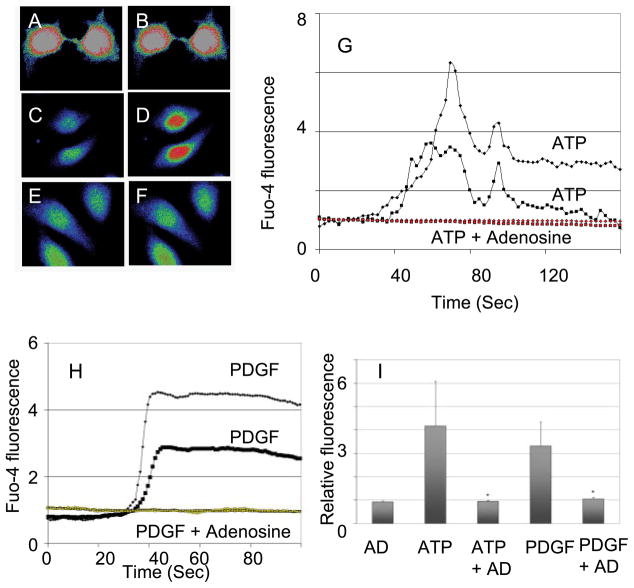
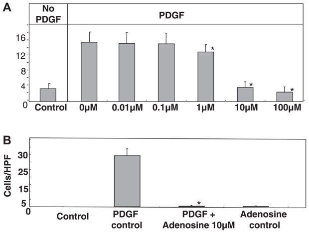
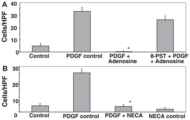
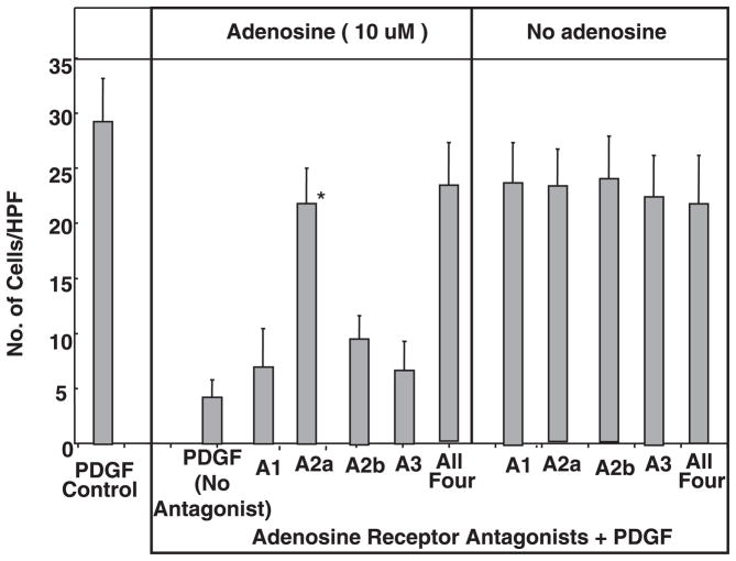
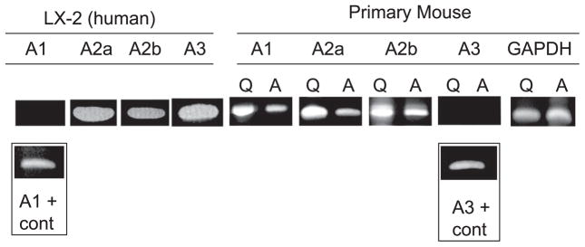
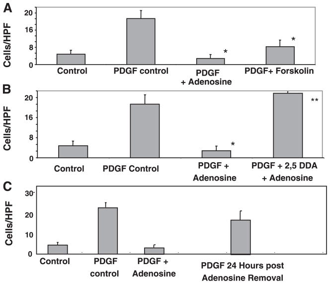
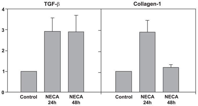
Adenosine is produced during cellular hypoxia and apoptosis, resulting in elevated tissue levels at sites of injury. Adenosine is also known to regulate a number of cellular responses to injury, but its role in hepatic stellate cell (HSC) biology and liver fibrosis is poorly understood. We tested the effect of adenosine on the cytosolic Ca2+ concentration, chemotaxis, and upregulation of activation markers in HSCs. We showed that adenosine did not induce an increase in the cytosolic Ca2+ concentration in LX-2 cells and, in addition, inhibited increases in the cytosolic Ca2+ concentration in response to ATP and PDGF. Using a Transwell system, we showed that adenosine strongly inhibited PDGF-induced HSC chemotaxis in a dose-dependent manner. This inhibition was mediated via the A(2a) receptor, was reversible, was reproduced by forskolin, and was blocked by the adenylate cyclase inhibitor 2,5-dideoxyadenosine. Adenosine also upregulated the production of TGF-beta and collagen I mRNA. In conclusion, adenosine reversibly inhibits Ca2+ fluxes and chemotaxis of HSCs and upregulates TGF-beta and collagen I mRNA. We propose that adenosine provides 1) a "stop" signal to HSCs when they reach sites of tissue injury with high adenosine concentrations and 2) stimulates transdifferentiation of HSCs by upregulating collagen and TGF-beta production.


Figures

Fig. 1
Fig. 1
Adenosine inhibits increase in cytosolic Ca2+ induced by ATP and PDGF. A: human LX-2 hepatic stellate cell (HSC) loaded with a Ca2+-sensing dye (fluorophore fluo-4 AM) before the addition of adenosine. B: same cell as in A at 60 s after the addition of adenosine (10 μM) with no change in cytosolic Ca2+. C: untreated LX-2 cell. D: same cell as in C at 60 s after the addition of ATP at 100 μM. E: LX-2 cell treated with adenosine (10 μM) for 10 min. F: same cell as in E at 60 s after the addition of ATP at 100 μM. G: representative plots of changes in cytosolic Ca2+ after treatment of the cells with ATP alone or ATP after cells had been exposed to adenosine (10 μM for 10 min). H: representative plots of changes in cytosolic Ca2+ after treatment of the cells with PDGF alone (10 ng/ml) or PDGF after cells had been exposed to adenosine (10 μM for 10 min). I: summary of relative changes in the fluorescence of fluorophore fluo-4 AM-loaded LX-2 cells after treatment with the listed agents. In all instances, when adenosine was used, it was at 10 μM and for 10 min before the addition of ATP or PDGF.
Fig. 2
Fig. 2
A: adenosine inhibited PDGF-induced chemotaxis in a dose-dependent manner. Human LX-2 HSCs (20,000 cells) were plated in the upper chamber of Transwell inserts with recombinant human PDGF-BB (10 ng/ml) as the chemoattractant in the lower chamber. Hematoxylin and eosin staining of the undersurface was done at the 24-h time point with migrating cells counted per high-power field (HPF). To test for the effect of adenosine on chemotaxis, cells were incubated with adenosine (at doses of 0.01, 0.1, 1, 10, and 100 μM) for 120 min before the addition of PDGF, and adenosine was maintained in the culture medium until the end of the 24-h study period (*P ≤ 0.05 vs. the PDGF control by Student’s t-test). B: primary mouse HSC chemotaxis was inhibited by adenosine. Primary mouse HSCs were isolated from B6 mice and cultured on plastic. Cells at day 5 were plated (20,000 cells) in the upper chamber of Transwell inserts, with PDGF-BB (10 ng/ml) as the chemoattractant in the lower chamber. Experiments were performed as in A with the addition of adenosine 120 min before the addition of PDGF (*P ≤ 0.05 vs. the PDGF control by Student’s t-test).
Fig. 3
Fig. 3
A: the pan-adenosine receptor antagonist 8-(p-sulfo-phenyl)-theophylline (8-SPT) blocked the ability of adenosine to inhibit PDGF-induced chemotaxis. Experiments were performed as in Fig. 2A with the addition of 8-SPT (10 μM) 15 min before the addition of adenosine. B: the nonhydrolysable adenosine agonist 5′-(N-ethylcarboxamido)adenosine (NECA) inhibited chemotaxis of LX-2 cells induced by PDGF. Experiments were performed as in Fig. 2A except that NECA (10 μM) was added 120 min before the addition of PDGF (*P ≤ 0.05 vs. the PDGF control by Student’s t-test).
Fig. 4
Fig. 4
Inhibition of PDGF (10 ng/ml)-mediated HSC chemotaxis by adenosine was reversed by preincubation with the A2a-specific receptor antagonist ZM-241835 at 1 μM [*P ≤ 0.05 vs. the PDGF (no antagonist) + adenosine (10 μM) control by Student’s t-test], whereas the A1 receptor antagonist 8-cyclopentyl-1,3-dipropylxanthine (DPCPX) at 10 nM, A2b receptor antagonist MRS-1706 at 10 nM, and the A3 receptor antagonist MRS-1523 at 5 μM had no effect. A similar antagonism of adenosine inhibition was seen with all four inhibitors combined at the above concentrations. Experiments were performed as in Fig. 3A with the addition of the receptor antagonists 15 min before the addition of adenosine.
Fig. 5
Fig. 5
Expression of adenosine receptor mRNA in LX-2 cells, quiescent primary mouse HSCs on day 1 of cell isolation (Q), and plastic activated primary mouse HSCs on day 5 (A). cDNA was prepared from and amplified with previously published primers and under previously published conditions. cDNA from human peripheral blood lymphocytes was used as positive control for human cells and the mouse brain for mouse primers, and gave a product for all four receptor subtypes. In LX-2 cells, mRNA for A2a, A2b, and A3 receptor subtypes was readily detected. In primary mouse HSCs, mRNA for A1, A2a, and A2b receptor subtypes was readily detected.
Fig. 6
Fig. 6
A: inhibition of HSC chemotaxis by adenosine (*P ≤ 0.05 vs. the PDGF control by Student’s t-test) was reproduced by the cAMP-inducing agent forskolin. Forskolin (100 μM) was added 30 min before the addition of PDGF, and the rest of the assay was performed as previously described (*P ≤ 0.05 vs. the PDGF control). B: inhibition of HSC chemotaxis by adenosine (*P ≤ 0.05 vs. the PDGF control) was blocked by the adenyl cyclase inhibitor 2,5-dideoxyadenosine (2,5-DDA). 2,5-DDA (100 μM) was added 30 min before the addition of adenosine (**P ≤ 0.05 vs. PDGF + adenosine). C: inhibition of PDGF-induced chemotaxis by adenosine was reversible. Adenosine inhibited PDGF-induced chemotaxis as shown in Fig. 2. Adenosine was removed from the media, and PDGF was added 24 h later. LX-2 cells were able to undergo chemotaxis in response to PDGF.
Fig. 7
Fig. 7
The adenosine receptor agonist NECA induced upregulation of TGF-β and collagen I mRNA. LX-2 cells were cultured in the presence or absence of NECA (10 μM), and cDNA was isolated 24 and 48 h later. Semiquantitative real-time PCR was performed using commercially available primer probe combinations. Results were standardized to GAPDH and are shown relative to untreated LX-2 cells. NECA induced upregulation of TGF-β at the 24- and 48-h time points (P ≤ 0.05 vs. the control) and induced upregulation of collagen I at 24 h (P ≤ 0.05 vs. the control).

References

    1. Aguirre-Ghiso JA, Estrada Y, Liu D, Ossowski L. ERK (MAPK) activity as a determinant of tumor growth and dormancy; regulation by p38 (SAPK) Cancer Res. 2003;63:1684–1695. - PubMed
    1. Albanis E, Friedman SL. Hepatic fibrosis. Pathogenesis and principles of therapy. Clin Liver Dis. 2001;5:315–334. - PubMed
    1. Bonner JC. Regulation of PDGF and its receptors in fibrotic diseases. Cytokine Growth Factor Rev. 2004;15:255–273. - PubMed
    1. Dawson AP. Calcium signalling: how do IP3 receptors work? Curr Biol. 1997;7:R544–R547. - PubMed
    1. Friedman SL. Mechanisms of disease: mechanisms of hepatic fibrosis and therapeutic implications. Nat Clin Pract Gastroenterol Hepatol. 2004;1:98–105. - PubMed

Publication types

MeSH terms