. 2006 Nov;116(11):3050-9.
doi: 10.1172/JCI28139. Epub 2006 Oct 19.

Toll-like receptor 4 deficiency causes pulmonary emphysema

Affiliations

Toll-like receptor 4 deficiency causes pulmonary emphysema

Xuchen Zhang et al. J Clin Invest. 2006 Nov.

Abstract

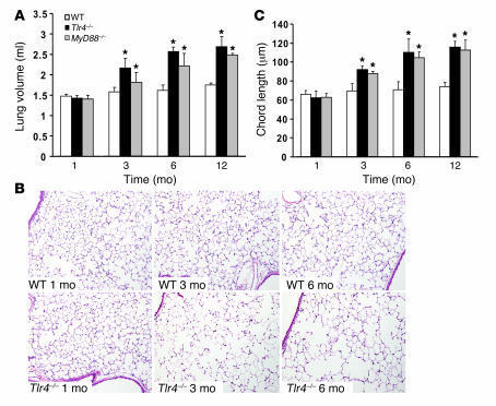
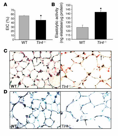
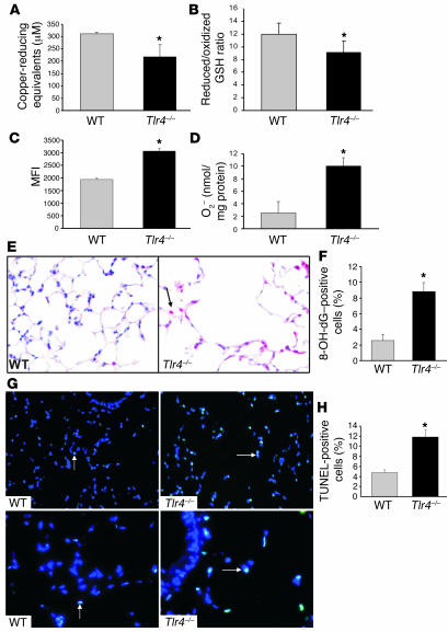
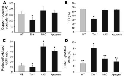
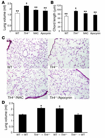
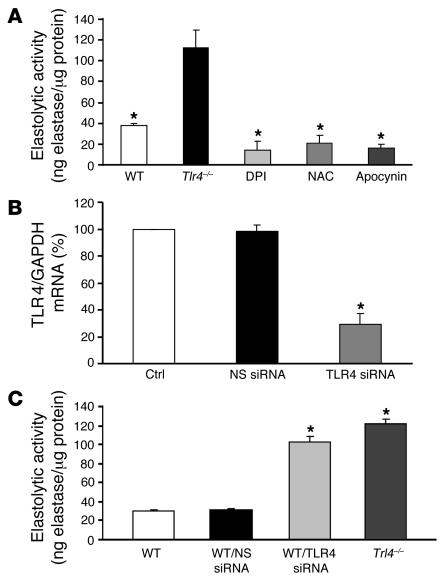
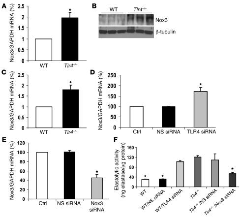
TLRs have been studied extensively in the context of pathogen challenges, yet their role in the unchallenged lung is unknown. Given their direct interface with the external environment, TLRs in the lungs are prime candidates to respond to air constituents, namely particulates and oxygen. The mechanism whereby the lung maintains structural integrity in the face of constant ambient exposures is essential to our understanding of lung disease. Emphysema is characterized by gradual loss of lung elasticity and irreversible airspace enlargement, usually in the later decades of life and after years of insult, most commonly cigarette smoke. Here we show Tlr4(-/-) mice exhibited emphysema as they aged. Adoptive transfer experiments revealed that TLR4 expression in lung structural cells was required for maintaining normal lung architecture. TLR4 deficiency led to the upregulation of what we believe to be a novel NADPH oxidase (Nox), Nox3, in lungs and endothelial cells, resulting in increased oxidant generation and elastolytic activity. Treatment of Tlr4(-/- )mice or endothelial cells with chemical NADPH inhibitors or Nox3 siRNA reversed the observed phenotype. Our data identify a role for TLR4 in maintaining constitutive lung integrity by modulating oxidant generation and provide insights into the development of emphysema.


Figures

Figure 1
Figure 1. Spontaneous pulmonary emphysema in Tlr4–/– mice.
(A) Lung volumes of WT, Tlr4–/–, and MyD88–/– mice. (B) Lung histology of WT and Tlr4–/– mice. Original magnification, ×100. (C) Mean linear chord length in WT, Tlr4–/–, and MyD88–/– mice (n = 5–7). Data are mean ± SEM. *P < 0.05 versus respective WT values.
Figure 2
Figure 2. Decreased EIC and increased elastin degradation in Tlr4–/– mice.
(A) EIC was detected in BAL of 3-month-old WT and Tlr4–/– mice (n = 5–6). (B) Elastolytic activity was assayed in lung lysates of 3-month-old WT and Tlr4–/– mice (n = 3). (C) Orcein staining for elastin in the elastic fibers of 3-month-old WT and Tlr4–/– mice. (D) Immunohistochemistry using an antibody directed against elastin in the elastic fibers of 3-month-old WT and Tlr4–/– mice. Arrows indicate representative elastin staining in the fibers (brown-black). Original magnification, ×400. Data are mean ± SEM. *P < 0.05 versus WT.
Figure 3
Figure 3. Decreased antioxidant capacity and increased oxidant burden in Tlr4–/– mice.
(A) Total antioxidant activity was measured in the BAL of 3-month-old WT and Tlr4–/– mice (n = 5–6). (B) The ratio of reduced GSH to oxidized GSH was detected in BAL of 3-month-old WT and Tlr4–/– mice (n = 6). (C) Quantitation of flow cytometric results from dihydroethidine staining in total lung cells isolated from 3-month-old WT and Tlr4–/– mice (n = 3). MFI, mean fluorescence intensity. (D) Detection of O2 production by cytochrome C reduction assays in total lung cells isolated from 3-month-old WT and Tlr4–/– mice (n = 3). (E) Representative DNA oxidation detection by 8-OH-dG immunohistochemical staining in the lungs of 3-month-old WT and Tlr4–/– mice. Arrow indicates positive red staining for 8-OH-dG. Original magnification, ×400. (F) Graphical quantitation of 8-OH-dG–positive cells expressed as % of total cells in lung sections (n = 3). (G) Representative merged images of lung sections stained with TUNEL (green) and DAPI stain (blue) from 3-month-old WT and Tlr4–/– mice. Arrows indicate representative TUNEL-positive cells (blue-green). Original magnification, ×200 (top panels); ×400 (bottom panels). (H) Graphical quantitation of TUNEL-positive cells expressed as percent of total cells in lung sections (n = 3). Data are mean ± SEM. *P < 0.05 versus WT.
Figure 4
Figure 4. Antioxidant administration restores the antioxidant activity, EIC, and cell survival in Tlr4–/– mice.
Tlr4–/– mice were fed NAC, apocynin, or vehicle-only water and sacrificed at 3 months of age to determine total antioxidant activity in BAL (A); EIC in BAL (B); reduced GSH/oxidized GSH ratio in BAL (C); and TUNEL-positive cells (expressed as percent of total cells) in lung sections (D). *P < 0.05 versus WT; **P < 0.05 versus Tlr4–/–. n = 4–5.
Figure 5
Figure 5. Antioxidant administration ameliorates alveolar enlargement in Tlr4–/– mice, whereas adoptive transfer of WT bone marrow cells has no effect.
(A) Lung volume. (B) Mean linear chord length. n = 4–5. (C) Representative lung histology by H&E. Original magnification, ×100. (D) Lung volume was measured 2 months after adoptive transfer of bone marrow cells from WT mice to WT mice (WT → WT), Tlr4–/– mice to Tlr4–/– mice (Tlr4–/–Tlr4–/–), WT mice to Tlr4–/– mice (WT → Tlr4–/–), and Tlr4–/– mice to WT mice (Tlr4–/– → WT). Data are mean ± SEM. *P < 0.05 versus WT or WT → WT; **P < 0.05 versus Tlr4–/–.
Figure 6
Figure 6. TLR4 deficiency leads to increased Nox-mediated elastolytic activity in MLECs.
(A) MLECs isolated from Tlr4–/– mice were treated with DPI (10 μM), NAC (100 μM), or apocynin (10 μM) for 24 hours, and elastolytic activity was assayed (n = 3). (B) TLR4 siRNA and nonspecific (NS) siRNA (80 nM) were transfected to WT MLECs, and TLR4 mRNA expression was analyzed by real-time RT-PCR (n = 3–5). Ctrl, untransfected control. (C) WT MLECs were transfected with nonspecific siRNA or TLR4 siRNA (80 nM), and elastolytic activity was assayed (n = 3). Data are mean ± SEM. *P < 0.05 versus Tlr4–/– (A), control and nonspecific siRNA (B), and WT and WT transfected with nonspecific siRNA (C).
Figure 7
Figure 7. TLR4 deficiency leads to increased Nox3 expression in lung and lung endothelial cells.
(A) Nox3 mRNA expression in 3-month-old WT and Tlr4–/– mouse lungs as detected by real-time RT-PCR (n = 3). (B) Nox3 protein expression in 3-month-old WT and Tlr4–/– mouse lungs (n = 3). (C) Nox3 mRNA expression in MLECs isolated from WT and Tlr4–/– mice as detected by real-time RT-PCR (n = 3). (D) Nox3 mRNA expression in WT MLECs transfected with nonspecific siRNA or TLR4 siRNA (80 nM) as detected by real-time RT-PCR compared with untransfected controls (n = 3). (E) Nox3 mRNA expression in WT MLECs transfected with nonspecific siRNA or Nox3 siRNA (80 nM) as detected by real time RT-PCR compared with untransfected controls (n = 3). (F) Elastolytic activity was detected in Tlr4–/– MLECs transfected with Nox3 siRNA or nonspecific siRNA and WT MLECs transfected with TLR4 siRNA or nonspecific siRNA (n = 3). Data are shown as mean ± SEM. *P < 0.05 versus WT (A and C), untransfected controls (D and E), and Tlr4–/– (F).

References

    1. Zhang X., et al. Cutting edge: TLR4 deficiency confers susceptibility to lethal oxidant lung injury. J. Immunol. 2005;175:4834–4838. - PubMed
    1. Qureshi S.T., et al. Inducible activation of TLR4 confers resistance to hyperoxia-induced pulmonary apoptosis. J. Immunol. 2006;176:4950–4958. - PubMed
    1. Jiang D., et al. Regulation of lung injury and repair by Toll-like receptors and hyaluronan. Nat. Med. 2005;11:1173–1179. - PubMed
    1. MacNee W. Pulmonary and systemic oxidant/antioxidant imbalance in chronic obstructive pulmonary disease. Proc. Am. Thorac. Soc. 2005;2:50–60. - PubMed
    1. Rennard S.I., Vestbo J. COPD: the dangerous underestimate of 15%. Lancet. 2006;367:1216–1219. - PubMed

Publication types

MeSH terms