. 2006 Dec 15;364(5):955-63.
doi: 10.1016/j.jmb.2006.09.075. Epub 2006 Oct 3.

Arrestin binding to calmodulin: a direct interaction between two ubiquitous signaling proteins

Affiliations

Arrestin binding to calmodulin: a direct interaction between two ubiquitous signaling proteins

Nan Wu et al. J Mol Biol. .

Abstract

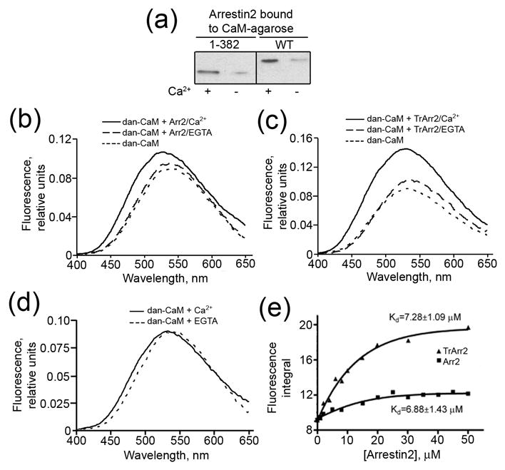
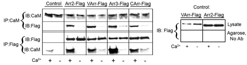
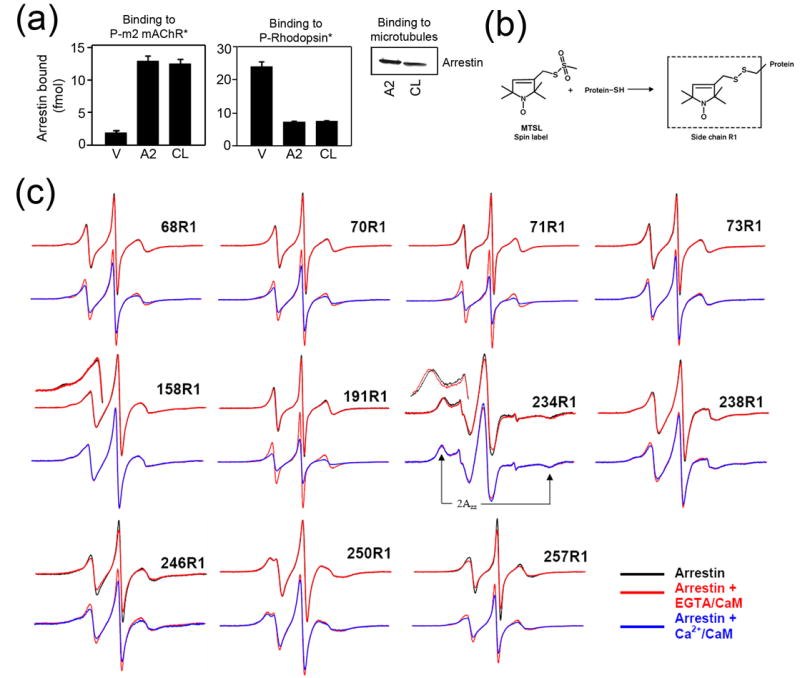
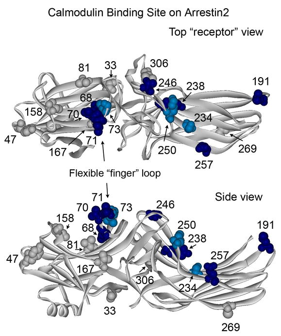
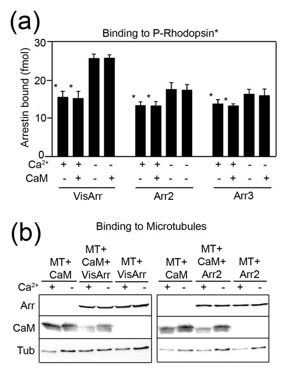
Arrestins serve as multi-functional regulators of G-protein coupled receptors, interacting with hundreds of different receptor subtypes and a variety of other signaling proteins. Here we identify calmodulin as a novel arrestin interaction partner using three independent methods in vitro and in cells. Arrestin preferentially binds calcium-loaded calmodulin with a Kd value of approximately 7 microM, which is within range of endogenous calmodulin concentrations. The calmodulin binding site is localized on the concave side of the C-domain and a loop in the center of the arrestin molecule, significantly overlapping with receptor and microtubule-binding sites. Using purified proteins, we found that arrestins sequester calmodulin, preventing its binding to microtubules. Nanomolar affinity of arrestins for their cognate receptors makes calmodulin an ineffective competitor for arrestin binding at relatively high receptor concentrations. The arrestin-calmodulin interaction likely regulates the localization of both proteins and their availability for other interaction partners.


Figures

Figure 1
Figure 1. Calcium-dependent interaction of arrestin2 with calmodulin
(a) Equal proportions of wild type (WT) and truncated (1-382) arrestin2 bound to CaM-agarose in the presence of 0.1mM CaCl2 or 5mM EGTA were run on SDS-PAGE and analyzed by Western blot with anti-arrestin antibody. “Empty” CNBr-activated Sepharose treated and blocked under the same conditions as CaM-agarose was used as a negative control. Non-specific arrestin binding was low (<2% of the input) and independent of Ca2+ (not shown). (bd) Representative spectra of dansyl-CaM (5μM) in the presence of 20μM WT (b), truncated (Tr) (c) arrestin2 (Arr2), and in the absence of arrestin2 (d) in buffer containing 0.1mM CaCl2 or 5mM EGTA. The average spectra of three experiments are shown. (e) The integrated area under the emission spectrum plotted vs the concentration of arrestin2. The data were fit to the Boltzmann one-site binding equation and the dissociation constant was calculated according to Bertrand et al as described in the Methods.
Figure 2
Figure 2. All four mammalian arrestins bind Ca2+/CaM in cells
The lysates of cells expressing HA-CaM and the indicated flag-tagged arrestins (or empty vector; control) were immunoprecipitated (IP) with rabbit anti-Flag or rabbit anti-calmodulin (CaM) antibodies as described in the Methods. Aliquots of the total lysate and immunoprecipitated proteins were run on 12.5% (calmodulin) or 10% (arrestin) SDS-PAGE and blotted (IB) for HA-CaM or Flag-arrestin. Non-specific binding of arrestins to Protein G agarose (in the absence of antibody (No Ab)), was very low and calcium-independent (right panel). Visual (VArr), arrestin2 (Arr2), arrestin3 (Arr3), and cone arrestin (CArr) were expressed at 96+30, 74+26, 53+9, and 73+21 pmol/mg protein, respectively, yielding intracellular concentrations that are somewhat higher than those found in mature neurons for non-visual arrestins and lower than those found in rod photoreceptors for visual arrestin ; . HA-CaM was expressed at a level equal to that of endogenous CaM and was used to facilitate immunoblotting. Representative results from 2–4 experiments are shown.
Figure 3
Figure 3. The localization of the calmodulin binding site on arrestin2 using site-directed spin labeling EPR spectroscopy
(a) Binding of visual (V), arrestin2 (A2), and cysless arrestin2 (CL) to the phosphorylated carbachol-activated m2 muscarinic receptor (P-m2 mAChR*) (left panel), light-activated phosphorylated rhodopsin (P-Rhodopsin*) (center panel), and microtubules (right panel) was performed as described in the Methods. Functionally cysless arrestin2 was identical to wild type arrestin2 in all cases. (b) The R1 side chain generated by reacting the arrestin cysteine mutants with the methanethiosulfonate (MTSL) nitroxide spin label reagent. (c) For each spin-labeled arrestin, normalized spectra in the absence (black) or presence of CaM + 1mM EGTA (red) are compared in the top row, and spectra in the presence of CaM + 1mM EGTA (red) and CaM + 0.1mM Ca2+ (blue) are compared in the bottom row. Portions of the overlaid spectra for 158R1 and 234R1 are magnified to better illustrate the spectral changes and the location of the hyperfine splitting (2Azz'). The spectrum of each arrestin mutant alone in solution or with 1mM EGTA or 0.1mM Ca2+ in the absence of calmodulin (black) were identical (spectra not shown). Only the spectra showing significant changes in the presence of calmodulin are presented.
Figure 4
Figure 4. Summary of the changes in spin label mobility induced by arrestin interaction with Ca2+/CaM
The magnitude of Ca2+/CaM-induced changes in spin label mobility (Fig.3) is color-coded on the arrestin2 crystal structure (PDB ID: 1G4R) as follows: light blue/dark blue, small and large changes in mobility, respectively; gray, no change (spectral data not shown for sites 33, 47, 81, 167, 269, and 306). Top panel: view from the “receptor binding” side of arrestin. Bottom panel: side view.
Figure 5
Figure 5. The effects of Ca2+/CaM on arrestin binding to the receptor and microtubules
(a) Binding of radiolabeled arrestins to light-activated phosphorylated rhodopsin (P-Rhodopsin*) was performed in the presence (+) or absence (−) of 1.3μM free Ca2+ and in the presence (+) or absence (−) of 10μM purified calmodulin (CaM) as described in the Methods. VisArr, visual arrestin; Arr2, arrestin2; Arr3, arrestin3. (b) Binding of purified arrestins (0.44μM), and/or purified calmodulin (0.44μM), to pre-polymerized taxol-stabilized microtubules (MT) (1.8μM of tubulin αβ-dimer) was performed in the presence (+) or absence (−) of 1.3μM free Ca2+ as described in the Methods. Representative Western blots of the microtubule pellet fraction are shown.

References

    1. Carman CV, Benovic JL. G-protein-coupled receptors: turn-ons and turn-offs. Curr Opin Neurobiol. 1998;8:335–344. - PubMed
    1. Lefkowitz RJ, Shenoy SK. Transduction of receptor signals by beta-arrestins. Science. 2005;308:512–517. - PubMed
    1. Gurevich VV, Gurevich EV. The new face of active receptor bound arrestin attracts new partners. Structure (Camb) 2003;11:1037–1042. - PubMed
    1. Gurevich VV, Gurevich EV. The molecular acrobatics of arrestin activation. TIPS. 2004;25:59–112. - PubMed
    1. Vishnivetskiy SA, Hosey MM, Benovic JL, Gurevich VV. Mapping the arrestin-receptor interface: structural elements responsible for receptor specificity of arrestin proteins. J Biol Chem. 2004;279:1262–1268. - PubMed

Publication types

LinkOut - more resources