. 2006 Nov 13;580(26):6062-8.
doi: 10.1016/j.febslet.2006.09.072. Epub 2006 Oct 16.

The Bysl gene product, bystin, is essential for survival of mouse embryos

Affiliations

The Bysl gene product, bystin, is essential for survival of mouse embryos

Rui Aoki et al. FEBS Lett. .

Abstract

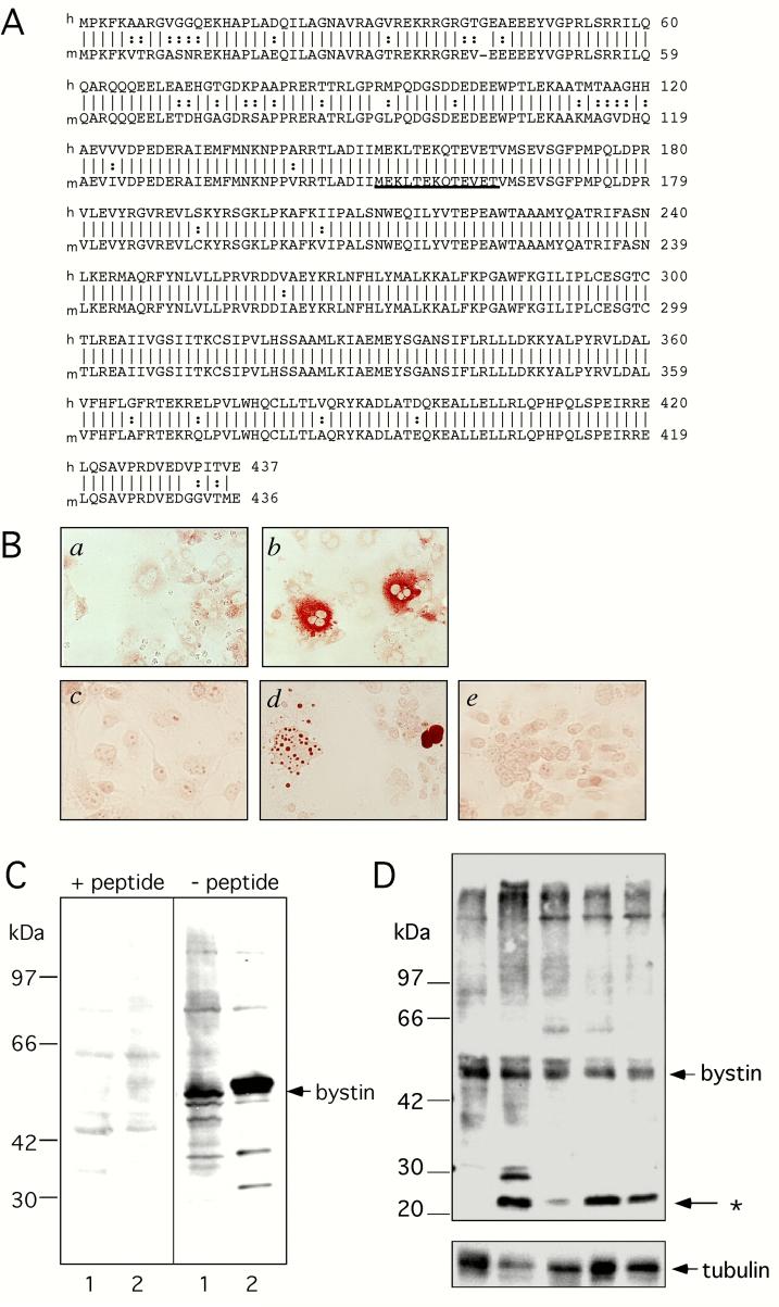
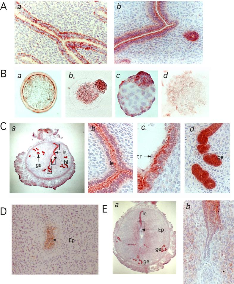
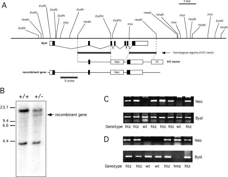
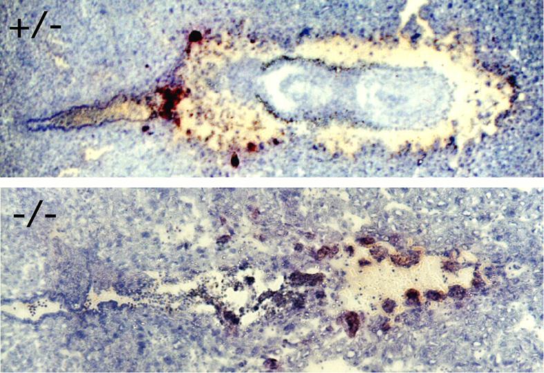
Human bystin is a cytoplasmic protein directly binding to trophinin, a cell adhesion molecule potentially involved in human embryo implantation. The present study shows that bystin is expressed in luminal and glandular epithelia in the mouse uterus at peri-implantation stages. In fertilized embryos, bystin was not seen until blastocyst stage. Bystin expression started during hatching and increased in expanded blastocyst. However, bystin apparently disappeared from the blastocyst during implantation. After implantation bystin re-appeared in the epiblast. Targeted disruption of the mouse bystin gene, Bysl, resulted in embryonic lethality shortly after implantation, indicating that bystin is essential for survival of mouse embryos.


Figures

Fig. 1
Fig. 1
Characterization of anti-bystin antibody by immunocytochemistry and Western blot. A, Peptide sequences of full-length human (upper) and mouse (lower) bystin proteins. The underlined sequence was used for antibody production. B. Immunohistochemistry of COS cells transfected with vector alone (a, c) or a vector encoding mouse bystin (b, d, e). Cells were reacted with an anti-bystin antibody with (e) or without (a-d) the peptide used as immunogen. C, Western blot analysis of FLAG-tagged human bystin. Each lane represents an untransfected (lane 1) and a transfected (lane 2) 293T cell lysate. The filter was reacted with an anti-bystin antibody with (left) or without (right) the epitope peptide. D. Western blot analysis of mouse uterus tissues. Lanes 1 and 2: non-pregnant. Lanes 3-5, pregnant day 3.5 (lane 3), day 4.5 (lane 4) and day 5.5 (lane 6).
Fig. 2
Fig. 2
Immunohistochemistry of mouse uteri and embryos before, during and after implantation. A, mouse uterus from non-pregnant (a) and 4.5 day pregnant (b) mice. B, Pre-implantation stage mouse embryos. Morulae (a), blastocysts during hatching (b), a hatched and expanded blastocyst (c), and a hatched and one day cultured blastocyst (d). C, Day 4.5 pregnant mouse uterus with implanting blastocyst. Bystin was seen in the uterine luminal epithelial cells (le) and glandular epithelial cells (ge), at low (a) and high (b-d) magnification. Note that in luminal epithelial cells, bystin localizes to the apical side (b). In luminal epithelial cells adjacent to the blastocyst, bystin localizes to the apical side, whereas trophectoderm cells do not show bystin signals (c). Glandular epithelia cells strongly express bystin, whereas no polarization of bystin staining is seen in these cells (d). D, Implanted embryo at the epiblast stage or at day 5.5. E. Implanted embryo and uterus at day 6.5. ge, glandular epithelia; le, luminal epithelia; bl, blastocyst; tr, trophectoderm; ep, epiblast.
Fig. 3
Fig. 3
Production of Bysl gene knockout mouse. A, Genomic DNA map of the Bysl gene, and the Bysl targeting vector. B, Southern blot of homologous recombinant ES clones. Genomic DNA digested by EcoRI was probed by the 5’ DNA probe shown in A. C, Genotyping by PCR for Neo gene and Bysl gene of genomic tail DNA from pups produced from the cross between Bysl (+/-) parents. D, Genotyping by PCR for Neo gene and Bysl gene of mouse embryos produced from Bysl(+/-) parents.
Fig. 4
Fig. 4
Histology of implanted E6.5 mouse embryos resulted from Bysl(+/-) parents. A pair of litter mates with each genotype Bysl(+/-) (upper) and Bysl(-/-) (lower) embryos is shown. Note that Bysl (-/-) embryo was degenerated. Tissue sections were stained with hematoxylin.

References

    1. Suzuki N, Zara J, Sato T, Ong E, Bakhiet N, Oshima RG, Watson KL, Fukuda MN. A novel cytoplasmic protein, bystin, interacts with trophinin, tastin and cytokeratin, and may be involved in trophinin mediated cell adhesion between trophoblast and endometrial epithelial cells. Proc. Natl. Acad. Sci. USA. 1998;95:5027–5032. - PMC - PubMed
    1. Fukuda MN, Sato T, Nakayama J, Klier G, Mikami M, Aoki D, Nozawa S. Trophinin and tastin, a novel cell adhesion molecule complex with potential involvement in embryo implantation. Genes Dev. 1995;9:1199–1210. - PubMed
    1. Fukuda MN, Nozawa S. Trophinin, tastin, and bystin: a complex mediating unique attachment between trophoblastic and endometrial epithelial cells at their respective apical cell membranes. Sem. Reprod. Endocrinol. 1999;17:229–34. - PubMed
    1. Suzuki N, Nakayama J, Shih IM, Aoki D, Nozawa S, Fukuda MN. Expression of trophinin, tastin, and bystin by trophoblast and endometrial cells in human placenta. Biol. Reprod. 1999;60:621–7. - PubMed
    1. Nakayama J, et al. Implantation-Dependent Expression of Trophinin by Maternal Fallopian Tube Epithelia during Tubal Pregnancies: Possible Role of Human Chorionic Gonadotrophin on Ectopic Pregnancy. Am. J. Pathol. 2003;163:2211–9. - PMC - PubMed

Publication types

Substances