Review
. 2006 Dec;9(6):595-601.
doi: 10.1016/j.mib.2006.10.007. Epub 2006 Oct 20.

Morphogenesis and cell cycle progression in Candida albicans

Affiliations
Review

Morphogenesis and cell cycle progression in Candida albicans

Judith Berman. Curr Opin Microbiol. 2006 Dec.

Abstract

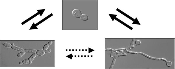
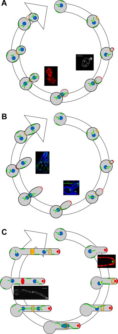
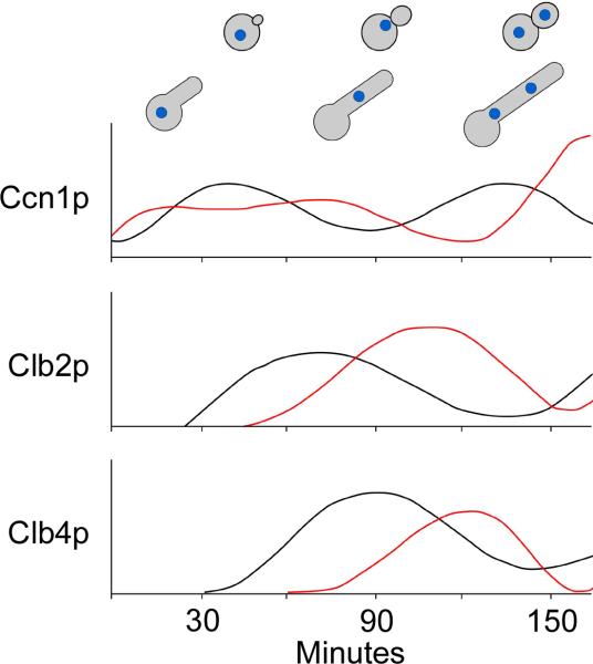
Candida albicans, an opportunistic human pathogen, displays three modes of growth: yeast, pseudohyphae and true hyphae, all of which differ both in morphology and in aspects of cell cycle progression. In particular, in hyphal cells, polarized growth becomes uncoupled from other cell cycle events. Yeast or pseudohyphae that undergo a cell cycle delay also exhibit polarized growth, independent of cell cycle progression. The Spitzenkörper, an organelle composed of vesicles associated with hyphal tips, directs continuous hyphal elongation in filamentous fungal species and also in C. albicans hyphae. A polarisome mediates cell cycle dependent growth in yeast and pseudohyphae. Regulation of morphogenesis and cell cycle progression is dependent upon specific cyclins, all of which affect morphogenesis and some of which function specifically in yeast or hyphal cells. Future work will probably focus on the cell cycle checkpoints involved in connecting morphogenesis to cell cycle progression.


Figures

Figure 1
Figure 1
Vegetative morphology of C. albicans cells. Yeast cells (top center) can form both pseudohyphae (lower left) and true hyphae (lower right). Switching between the pseudohyphal and hyphal morphologies is less frequent.
Figure 2
Figure 2
Models for cell cycle progression in yeast, pseudohyphal and hyphal cells. A. Yeast cells traverse START by forming a septin ring (orange), initiating bud emergence directed by a polarisome (red crescent) and duplicating the spindle pole body (yellow). Growth becomes less polarized as sites of growth (red) become distributed around the bud. In G2 phase the nucleus (blue) moves to the neck assisted by astral microtubule (green) sliding along the cortex and, at anaphase, divides across the neck. At telophase, the spindle disassembles, growth is focused at the neck, the septin ring splits into two and then each ring disappears prior to appearance of the next ring in G1. Right inset, polarisome protein Mlc1p-YFP localizes to the tip during early bud growth. Left inset, delocalized actin (red) patches reflect isotropic growth. B. Pseudohyphal cells have similar features to yeast cells with a few exceptions: the polarisome persists for longer and cells spend more time in G2 phase, becoming similar in size to mother cells; cells do not separate following cytokinesis. As in yeast cells, sites of growth are cell cycle dependent, leaving the tip and focusing at the bud neck prior to cytokinesis. Right inset, Mlc1p-GFP (green) appears at the tips of small and larger buds. Left inset, at cytokinesis, Mlc1p-GFP disappears from bud tip and localizes to the neck. C. Upon induction of hyphal growth from a yeast cell, the Spitzenkörper (red circle) directs germ tube evagination, which persists throughout the cell cycle and initiates prior to START. A polarisome is also present at hyphal tips. Nuclei migrate to and divide across the presumptum, and the septin ring persists into the next cell cycle. Right inset, photomicrograph of Spitzenkörper protein Mlc1p-YFP (green); cell surface is labeled with Texas-red conjugated to Concanavalin A. Left inset, during cytokinesis Mlc1p-YFP remains at the growing tip and also appears at the septum.
Figure 3
Figure 3
Cell cycle progression and cyclin levels differ in yeast and hyphae. G1-phase yeast daughter cells were synchronized by elutriation and then released into yeast (30°C) or hyphal (37°C, 5% serum) growth conditions. Cell morphology and levels of G1 cyclin Ccn1p, and B-cyclins Clb2p and Clb4p were followed using epitope tagged proteins. Ccn1p levels persisted longer and B-cyclins appeared later in hyphae, relative to yeast. Adapted from Bensen et al. 2005.
Figure 4/Box 1
Figure 4/Box 1
List of cell cycle conditions and mutants that cause changes in morphogenesis. Gene/conditions are ordered by approximate cell cycle stage at which arrest/delay occurs. Essential genes (asterisk) are terminally arrested. G1 arrested cells tend to be more hyphal-like, S/G2 and M arrests tend to be polarized pseudohyphal-like.

References

    1. Gow N, Brown A, Odds F. Fungal morphogenesis and host invasion. Curr Opin Microbiol. 2002;5:366. - PubMed
    1. Saville SP, Lazzell AL, Monteagudo C, Lopez-Ribot JL. Engineered control of cell morphology in vivo reveals distinct roles for yeast and filamentous frms of Candida albicans during infection. Eukaryot Cell. 2003;2:1053–1060. - PMC - PubMed
    1. Wightman R, Bates S, Amornrrattanapan P, Sudbery P. In Candida albicans, the Nim1 kinases Gin4 and Hsl1 negatively regulate pseudohypha formation and Gin4 also controls septin organization. J Cell Biol. 2004;164:581–591. - PMC - PubMed
    2. •This paper describes kinases that act at the septin ring and affect morphogenesis. It also shows that gin4Δ/Δ cells, which are pseudohyphal, cannot form hyphae, while Gin4p-depleted cells, which start in the yeast form, can form hyphae.

    1. Lew DJ, Reed SI. Cell cycle control of morphogenesis in budding yeast. Curr Opin Genet Dev. 1995;5:17–23. - PubMed
    1. Crampin H, Finley KR, Gerami-Nejad M, Court H, Gale CA, Berman J, Sudbery PE. Candida albicans hyphae have a Spitzenkörper that is distinct from the polarisome found in yeast and pseudohyphae. J. Cell Sci. 2005;118:2935–2947. - PubMed
    2. ••This manuscript demonstrates that C. albicans hyphae have a Spitzenörper in addition to a polarisome and that it persists throughout hyphal growth. It shows that components of polarisomes are found primarily in the Spitzenkörper (Mlc1p and Bni1p), while others (Spa2p, Bud6p and Cdc43p) are primarily found in the polarisome.

Publication types

MeSH terms

LinkOut - more resources