. 2006 Dec 1;456(1):93-7.
doi: 10.1016/j.abb.2006.09.024. Epub 2006 Oct 10.

Interaction between glycogenin and glycogen synthase

Affiliations

Interaction between glycogenin and glycogen synthase

Alexander V Skurat et al. Arch Biochem Biophys. .

Abstract

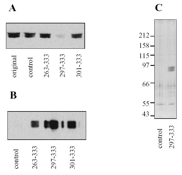
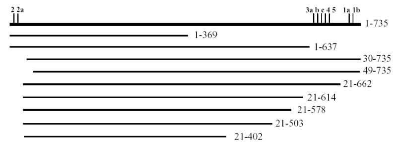
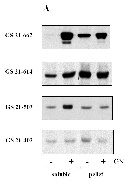
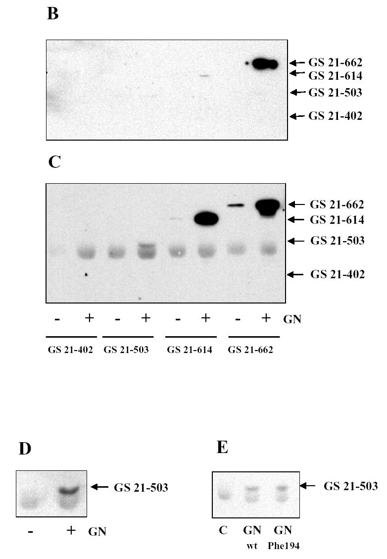
Glycogen synthase plays a key role in regulating glycogen metabolism. In a search for regulators of glycogen synthase, a yeast two-hybrid study was performed. Two glycogen synthase-interacting proteins were identified in human skeletal muscle, glycogenin-1, and nebulin. The interaction with glycogenin was found to be mediated by the region of glycogenin which contains the 33 COOH-terminal amino acid residues. The regions in glycogen synthase containing both NH2- and COOH-terminal phosphorylation sites are not involved in the interaction. The core segment of glycogen synthase from Glu21 to Gly503 does not bind COOH-terminal fragment of glycogenin. However, this region of glycogen synthase binds full-length glycogenin indicating that glycogenin contains at least one additional interacting site for glycogen synthase besides the COOH-terminus. We demonstrate that the COOH-terminal fragment of glycogenin can be used as an effective high affinity reagent for the purification of glycogen synthase from skeletal muscle and liver.


Figures

Figure 1
Figure 1. Interaction of COOH-terminal fragments of glycogenin with mouse muscle glycogen synthase
Mouse skeletal muscle was homogenized, centrifuged and supernatant (original) prepared. The recombinant GST-fusion proteins with C-terminal parts of glycogenin (amino acids 263–333, 297–333 and 301–333) or native GST (control) were added. GST-fusion proteins were precipitated by adding glutathione-agarose, and the resulting supernatants (A) and beads (B) were analyzed for the presence of glycogen synthase using immunoblotting. Glycogen synthase purified with GST-GN297-333 was eluted from beads by glutathione and analyzed by silver staining (C). The numbers to the left show the molecular masses and migration of protein standards.
Figure 2
Figure 2. Schematic representation of wild type and truncated glycogen synthase
Glycogen synthase protein is shown as horizontal line; the vertical tick marks indicate phosphorylation sites. The numbers to the left indicate the first and the last amino acids in the protein sequence of wild type (1–735) and truncated glycogen synthase.
Figure 3
Figure 3. Interaction of glycogenin and glycogen synthase expressed in COS cells
Glycogen synthase mutants with an NH2-terminal myc epitope tag were expressed in COS cells with (+) or without (−) glycogenin (GN). Proteins in the soluble and pellet fractions of COS cells were analyzed by immunoblotting (A). Glycogen synthase mutants were pulled down from the soluble fraction of COS cells using GST-GN297-333 (B) or immunoprecipitated with anti-glycogenin antibody (C) and detected by immunoblotting. To better separate glycogen synthase GS 21-503 from the immunoglobulin band, SDS-PAGE was performed in a gel with lower percentage of acrylamide (D). The glycogen synthase GS 21-503 was co-expressed with wild type or the Phe194 mutant of glycogenin followed by immunoprecipitation with anti-glycogenin antibodies and immunoblotting (E). All immunoblots were performed with anti-myc antibodies.

References

    1. Roach PJ, Skurat AV, Harris RA. Regulation of glycogen metabolism. In: Jefferson LS, Cherrington AD, editors. Handbook of Physiology, Vol. 2: The endocrine pancreas and regulation of metabolism. Oxford University Press; New York: 2001. pp. 609–647.
    1. Cohen P. Muscle Glycogen Synthase. The Enzymes. 1986;17:461–497.
    1. Roach PJ. Control of glycogen synthase by hierarchal protein phosphorylation. FASEB J. 1991;4:2961–2968. - PubMed
    1. Skurat AV, Roach PJ. Regulation of glycogen synthesis. In: LeRoith D, Olefsky JM, Taylor SI, editors. Diabetes Mellitus: A Fundamental and Clinical Text. third edition. Lippincott-Raven Publishers; Philadelphia: 2004. pp. 317–334.
    1. Cheng C, Mu J, Farkas I, Huang D, Goebl MG, Roach PJ. Requirement of the self-glucosylating initiator proteins Glg1p and Glg2p for glycogen accumulation in Saccharomyces cerevisiae. Mol Cell Biol. 1995;15:6632–6640. - PMC - PubMed

Publication types

MeSH terms