. 2006 Nov 1;177(9):6215-26.
doi: 10.4049/jimmunol.177.9.6215.

Regulatory T cells dampen pulmonary inflammation and lung injury in an animal model of pneumocystis pneumonia

Affiliations

Regulatory T cells dampen pulmonary inflammation and lung injury in an animal model of pneumocystis pneumonia

Laura McKinley et al. J Immunol. .

Abstract

CD4+CD25+FoxP3+ regulatory T cells are decreased in patients infected with HIV and have been shown to be critical in mediating Ag tolerance in the lung. Because a subset of Pneumocystis-infected individuals develop substantial lung injury, which can be modeled in immune reconstituted scid mice, we used mouse models of Pneumocystis carinii to investigate the role of regulatory T cells in opportunistic infection and immune reconstitution. In this study, we show that CD4+CD25+FoxP3+ cells are part of the host response to Pneumocystis in CD4+ T cell-intact mice. Moreover, lung injury and proinflammatory Th1 and Th2 cytokine levels in the bronchoalveolar lavage fluid and lung homogenate were increased following CD4+CD25- immune reconstitution in Pneumocystis-infected SCID mice but not in CD4+CD25+ T cell-reconstituted animals. The ability of CD4+CD25+ T cells to control inflammation and injury during the course of Pneumocystis was confirmed by treatment of wild-type C57BL/6 mice with anti-CD25 mAb. These data show that CD4+CD25+ T cells control pulmonary inflammation and lung injury associated with Pneumocystis infection both in the setting of immune reconstitution as well as new acquisition of infection.


Figures
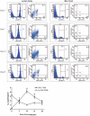
FIGURE 1
FIGURE 1
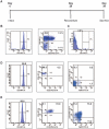
Trafficking of CD4+CD25+FoxP3+ T cells to the lung following Pneumocystis infection. Representative histograms from C57BL/6 mice infected with Pneumocystis for the indicated time. Day 0 animals were sacrificed immediately after inoculation. Surface staining of CD25 and intracellular FoxP3 expression are back-gated on the CD4+ T cell population. The data indicate there is a rise in the percentage of CD4+FoxP3+ in BAL fluid peaking at day 14 postinfection as LN regulatory cells decline. All data are reported as mean ± SEM. Day 0, n = 3; day 7, n = 6; day 14, n = 9; days 21 and 28, n = 5. *, p < 0.05 compared with LN day 0; #, p < 0.05 compared with BAL fluid day 0.
FIGURE 2
FIGURE 2
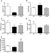
Assessment of pulmonary responses 14 days after Pneumocystis infection in WT mice treated with anti-CD4 or anti-CD25. A, Total BAL fluid cells. B, Organism burden determined by TaqMan PCR reported as copy number/whole lung. C, BAL fluid LDH. D, BAL fluid total protein. E, BAL fluid granzyme B. All data are reported as mean ± SEM for n = 7−9. *, p < 0.05 compared with WT. #, p < 0.05 compared with anti-CD4-treated animals.
FIGURE 3
FIGURE 3
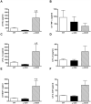
Treatment with CD25 mAb exacerbates pulmonary inflammation in Pneumocystis-infected animals. Lung homogenate levels of KC (A), TNF-α (B), IL-1β (C), IL-6 (D), MCP-1 (E), and G-CSF (F). All data are reported as mean ± SEM for n = 7−9. *, p < 0.05 compared with WT. #, p < 0.05 compared with anti-CD4-treated animals.
FIGURE 4
FIGURE 4
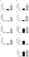
Treatment with anti-CD25 results in an exacerbated Th2 phenotype in the lungs of Pneumocystis-infected mice. A, BAL IL-4. B, Lung homogenate IL-4. C, BAL IL-5. D, Lung homogenate IL-5. E, BAL IL-13. F, Lung homogenate IL-13. G, BAL IFN-γ. H, Lung homogenate IFN-γ. I, Lung homogenate IL-17. BAL IL-17 was below detection. All data are reported as mean ± SEM for n = 7−9. *, p < 0.05 compared with WT. #, p < 0.05 compared with anti-CD4-treated animals.
FIGURE 5
FIGURE 5
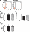
Secretion of IL-10 and TGF-β in the lung are not altered by treatment with anti-CD25. A, Representative histogram of BAL fluid cells stained for cell surface TGF-β and intracellular FoxP3 from control Ab-treated animals (left panel) or anti-CD25 mAb-treated animals (right panel). B, Active lung homogenate TGF-β. C, Active BAL fluid TGF-β. D, Lung homogenate IL-10. BAL fluid IL-10 was below detection. All data are reported as mean ± SEM for n = 7−9. *, p < 0.05 compared with WT. #, p < 0.05 compared with anti-CD4-treated animals.
FIGURE 6
FIGURE 6
SCID reconstitution model of Pneumocystis. A, SCID mice were inoculated on day 0 with 2 × 10⁵ Pneumocystis cysts/mouse intratracheally. On day 28, mice were reconstituted with 3 × 10⁵ WT cells as indicated; animals were sacrificed 11 days postreconstitution (day 39). Representative histograms of cell populations used for adoptive transfer. B, WT splenocytes were enriched for CD4 by negative selection. The starting cell population was consistently >90% CD4+; 5–10% of these cells coexpressed CD25. C, CD4 cells are those left after negative selection of CD4; <1% of this population expressed CD4. D, Purity of CD4+CD25 was >99%, and almost none of these cells expressed FoxP3. E, The majority of CD4+CD25+cells express intracellular FoxP3.
FIGURE 7
FIGURE 7
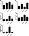
Reconstitution of Pneumocystis-infected SCID mice with CD4+CD25 T cells leads to significant lung injury. A, Total BAL fluid cells. B, Organism burden determined by TaqMan PCR reported as copy number/whole lung. C, BAL fluid LDH. D, BAL fluid total protein. E, BAL fluid granzyme B. All data are reported as mean ± SEM for n = 9−11. *, p < 0.05 compared with NR.
FIGURE 8
FIGURE 8
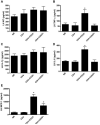
Hyperinflammatory profile in the lungs of Pneumocystis-infected mice reconstituted with CD4+CD25 T cells but not CD4 cells. Lung homogenate levels of KC (A), TNF-α (B), IL-1β (C), IL-6 (D), MCP-1 (E), and G-CSF (F). All data are reported as mean ± SEM for n = 9−11. *, p < 0.05 compared with NR.
FIGURE 9
FIGURE 9
Reconstitution with CD25 cells results in a mixed Th cytokine profile. A, Lung homogenate IL-5. B, Lung homogenate IL-13. C, Lung homogenate IFN-γ. D, Lung homogenate IL-17. BAL cytokine levels were below detection, as was lung homogenate IL-4. All data are reported as mean ± SEM for n = 9–11. *, p < 0.05 compared with NR.
FIGURE 10
FIGURE 10
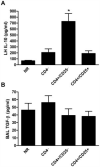
Reconstitution of infected SCID mice with regulatory T cells does not alter IL-10 or TGF-β secretion. BAL fluid IL-10 was below detection. A, Lung homogenate IL-10. B, Active BAL fluid TGF-β. All data are reported as mean ± SEM for n = 9−11. *, p < 0.05 compared with NR.

References

    1. Morris A, Lundgren JD, Masur H, Walzer PD, Hanson DL, Frederick T, Huang L, Beard CB, Kaplan JE. Current epidemiology of Pneumocystis pneumonia. Emerging Infect. Dis. 2004;10:1713–1720. - PMC - PubMed
    1. Phair J, Munoz A, Detels R, Kaslow R, Rinaldo C, Saah A. The risk of Pneumocystis carinii pneumonia among men infected with human immunodeficiency virus type 1: Multicenter AIDS Cohort Study Group. N. Engl. J. Med. 1990;322:161–165. - PubMed
    1. Stansell JD, Osmond DH, Charlebois E, LaVange L, Wallace JM, Alexander BV, Glassroth J, Kvale PA, Rosen MJ, Reichman LB, et al. Predictors of Pneumocystis carinii pneumonia in HIV-infected persons: pulmonary complications of HIV Infection Study Group. Am. J. Respir. Crit. Care Med. 1997;155:60–66. - PubMed
    1. Shellito J, Suzara VV, Blumenfeld W, Beck JM, Steger HJ, Ermak TH. A new model of Pneumocystis carinii infection in mice selectively depleted of helper T lymphocytes. J. Clin. Invest. 1990;85:1686–1693. - PMC - PubMed
    1. Harmsen AG, Stankiewicz M. Requirement for CD4+ cells in resistance to Pneumocystis carinii pneumonia in mice. Exp. Med. 1990;172:937–945. - PMC - PubMed

Publication types

MeSH terms