. 2006 Nov 1;177(9):6379-87.
doi: 10.4049/jimmunol.177.9.6379.

VCAM-1 signals activate endothelial cell protein kinase Calpha via oxidation

Affiliations

VCAM-1 signals activate endothelial cell protein kinase Calpha via oxidation

Hiam Abdala-Valencia et al. J Immunol. .

Abstract

Lymphocyte binding to VCAM-1 activates endothelial cell NADPH oxidase, resulting in the generation of 1 muM H(2)O(2). This is required for VCAM-1-dependent lymphocyte migration. In this study, we identified a role for protein kinase Calpha (PKCalpha) in VCAM-1 signal transduction in human and mouse endothelial cells. VCAM-1-dependent spleen cell migration under 2 dynes/cm(2) laminar flow was blocked by pretreatment of endothelial cells with dominant-negative PKCalpha or the PKCalpha inhibitors, Rö-32-0432 or Gö-6976. Phosphorylation of PKCalpha(Thr638), an autophosphorylation site indicating enzyme activity, was increased by Ab cross-linking of VCAM-1 on endothelial cells or by the exogenous addition of 1 muM H(2)O(2). The anti-VCAM-1-stimulated phosphorylation of PKCalpha(Thr638) was blocked by scavenging of H(2)O(2) and by inhibition of NADPH oxidase. Furthermore, anti-VCAM-1 signaling induced the oxidation of endothelial cell PKCalpha. Oxidized PKCalpha is a transiently active form of PKCalpha that is diacylglycerol independent. This oxidation was blocked by inhibition of NADPH oxidase. In summary, VCAM-1 activation of endothelial cell NADPH oxidase induces transient PKCalpha activation that is necessary for VCAM-1-dependent transendothelial cell migration.


Conflict of interest statement

Disclosures

The authors have no financial conflict of interest.

Figures
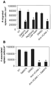
FIGURE 1
FIGURE 1
Inhibition of endothelial cell PKCα blocks VCAM-1-dependent spleen cell migration. A, Confluent monolayers of mHEVa cells were nontreated (NT) or treated for 20 min with the PKCα-selective inhibitors 2.3 nM Gö-6976 and 100 nM Rö-32-0432 and then washed five times. In addition, medium from the last washes was added to nontreated spleen cells to ensure that the inhibitor was sufficiently removed. Spleen cells were added to the endothelial monolayer in the presence or absence of anti-VCAM-1 (14 μg/cm2 endothelial cell monolayer) at 2 dynes/cm2, allowed to briefly settle to mediate cell contact and then exposed to 2 dynes/cm2 laminar flow for 15 min. Cells were washed and fixed in 3% paraformaldehyde for 1 h. Spleen cell migration was examined by phase contrast microscopy (28). Nonmigrated spleen cells are phase-light and migrated spleen cells appear as phase dark (28). The cells that migrated are >88% lymphocytes (10). Gö-6976 and Rö-32-0432 had no affect on cell viability, as determined by trypan blue exclusion (data not shown). Data are from three to five experiments.*, p < 0.05 compared with nontreated, DMSO treated, and last washes. B, Confluent monolayers of mHEVa cells were treated as in A with the inhibitors indicated, 2 dynes/cm2 laminar flow was applied for 3 min and then the cells were washed and fixed. The total number of spleen cells associated with the endothelial monolayer was determined. Data are from three experiments.*, p < 0.05 compared with cells without anti-VCAM-1 Abs.
FIGURE 2
FIGURE 2
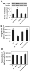
DN PKCα blocks VCAM-1-dependent spleen cell migration. mHEVa were grown to 90% confluence and transfected with 1 μg of vector with DN PKCα or vector pCMV for 3.5 h, washed, and cultured for 24 h. A, The cells were examined for total PKCα expression by Western blot. B, Spleen cells were added to the endothelial cell monolayer and the coculture was exposed to 2 dynes/cm2 laminar flow for 15 min as in Fig. 1. Cells were washed and fixed in 3% paraformaldehyde for 1 h. Spleen cell migration was examined by phase contrast microscopy. C, Spleen cells were added to the endothelial cell monolayer and the coculture was exposed to 2 dynes/cm2 laminar flow for 3 min as in Fig. 1. Cells were washed and fixed in 3% paraformaldehyde for 1 h. Total number of spleen cells associated with the endothelial cells was examined by microscopy. The DN PKCα had no affect on cell viability, as determined by trypan blue exclusion (data not shown). Data are from three experiments.*, p < 0.05 compared with nontreated.
FIGURE 3
FIGURE 3
Anti-VCAM-1 stimulation induces an increase in phosphorylation of PKCα Thr638 that is dependent on ROS generation. Confluent monolayers of A–C mHEVa cells and D TNF-α (1 ng/ml)-treated HMEC-L cells in 12-well plates were nontreated (NT) or incubated for 30 min with apocynin (4 mM), with catalase (5000 U/ml), or with DPI (5 μM). These endothelial cells were stimulated with 27 μg/ml anti-VCAM-1 Ab or control Ab plus 15 μg/ml of a secondary Ab for (A) 5–60 min or (B–D) 10 min. In D, the HMEC-L cells were also stimulated with 1 μM H2O2 for 10 min. Upper micrographs in each panel are representative Western blots using rabbit anti-phospho PKCα Thr638 (1/1000), rabbit anti-PKCα (1/100), or anti-β-actin (1/5000) followed by HRP-conjugated donkey anti-rabbit secondary Ab (1/4000) and ECL detection. The phosphorylation status of PKCα Thr638 is presented as the fold increase in the ratio of the relative intensity of phospho-PKCα Thr638/the relative intensity of the loading control (total PKCα or β-actin). The apocynin, catalase, DPI, and H2O2 had no effect on endothelial cell viability as determined by trypan blue exclusion (data not shown). Data presented are the mean ± SE from three experiments.*, p < 0.05 compared with NT, isotype Ab, or inhibitor-treated cells.
FIGURE 4
FIGURE 4
Exogenous 1 μM H2O2 activates endothelial cell-associated PKCα. A, mHEVa cells were incubated with low concentrations of exogenous H2O2 (0.1, 1, and 5 μM) for 10 min. Phospho-PKCα Thr638 was examined by Western blot and analyzed as described in Fig. 3.*, p < 0.05 as compared with 0, 0.1 and 5 μM H2O2.**, p < 0.05 as compared with 0 and 0.1 μM H2O2. B, mHEVa cells were incubated with 1 μM H2O2 for 1, 5, 10, or 15 min.*, p < 0.05 as compared with 1, 5, and 15 min.**, p < 0.05. A significant decrease compared with 10 min. Data presented are the mean ± SE from three experiments.
FIGURE 5
FIGURE 5
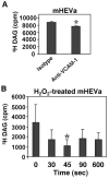
Anti-VCAM-1 and H2O2 stimulation does not increase endothelial cell DAG. Phospholipase C activity was assessed by measuring the production of [3H]DAG by mHEV cells prelabeled with (A) 1.5 μCi/well or (B) 0.5 μCi/well [3H]arachidonic acid for 48 h. A, Endothelial cells were stimulated with 27 μg/ml rat anti-mouse VCAM-1 Ab plus 15 μg/ml goat anti-rat IgG Ab for 10 min. B, Endothelial cells were stimulated with 1 μM H2O2 for 30 – 600 s.*, p < 0.05. Data presented are the mean ± SE from three experiments.
FIGURE 6
FIGURE 6
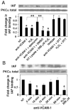
Anti-VCAM-1 and H2O2 induces oxidation of PKCα. mHEVa cells were treated with apocynin and then treated with anti-VCAM-1 or control Ab for 10 min as described in Fig. 3. A, The lysates were nontreated or reduced with 10 mM DTT for 10 min. As a positive control for oxidation, a lysate from NT cells was oxidized with 200 μM H2O2 for 20 min. The lysates were then examined for cysteine oxidation status by determining reactivity with 10 μM IAF (20 min), which reacts with nonoxidized cysteines. PKCα was immunoprecipitated and examined by Western blot using anti-fluorescein or anti-total PKCα Abs (1/1000). There was no change in total PKCα. B, mHEVa cells were stimulated for the time indicated with anti-VCAM-1 and examined for IAF reactivity as described in A. Data presented are the mean ± SD from three experiments.*, p < 0.05 as compared with NT.**, p < 0.05 for the comparison indicated.

References

    1. Iiyama K, Hajra L, Iiyama M, Li H, DiChiara M, Medoff BD, Cybulsky MI. Patterns of vascular cell adhesion molecule-1 and inter-cellular adhesion molecule-1 expression in rabbit and mouse atherosclerotic lesions and at sites predisposed to lesion formation. Circ Res. 1999;85:199–207. - PubMed
    1. Mueller JP, Evans MJ, Cofiell R, Rother RP, Matis LA, Elliott EA. Porcine vascular cell adhesion molecule (VCAM) mediates endothelial cell adhesion to human T cells: development of blocking antibodies specific for porcine VCAM. Transplantation. 1995;60:1299–1306. - PubMed
    1. Chin JE, Hatfield CA, Winterrowd GE, Brashler JR, Vonderfecht SL, Fidler SF, Griffin RL, Kolbasa KP, Krzesicki RF, Sly LM, et al. Airway recruitment of leukocytes in mice is dependent on α4-integrins and vascular cell adhesion molecule-1. Am J Physiol. 1997;272:L219–L229. - PubMed
    1. Baron JL, Madri JA, Ruddle NH, Hashim G, Janeway CA., Jr Surface expression of α4 integrin by CD4 T cells is required for their entry into brain parenchyma. J Exp Med. 1993;177:57–68. - PMC - PubMed
    1. Gurtner GC, Davis V, Li H, McCoy MJ, Sharpe A, Cybulsky MI. Targeted disruption of the murine VCAM1 gene: essential role of VCAM-1 in chorioallantoic fusion and placentation. Genes Dev. 1995;9:1–14. - PubMed

Publication types

MeSH terms