. 2006 Nov 1;177(9):6548-59.
doi: 10.4049/jimmunol.177.9.6548.

Gene transfer of tumor-reactive TCR confers both high avidity and tumor reactivity to nonreactive peripheral blood mononuclear cells and tumor-infiltrating lymphocytes

Affiliations

Gene transfer of tumor-reactive TCR confers both high avidity and tumor reactivity to nonreactive peripheral blood mononuclear cells and tumor-infiltrating lymphocytes

Laura A Johnson et al. J Immunol. .

Abstract

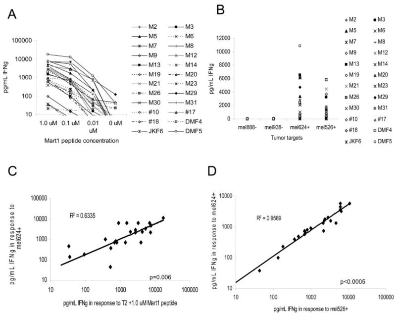
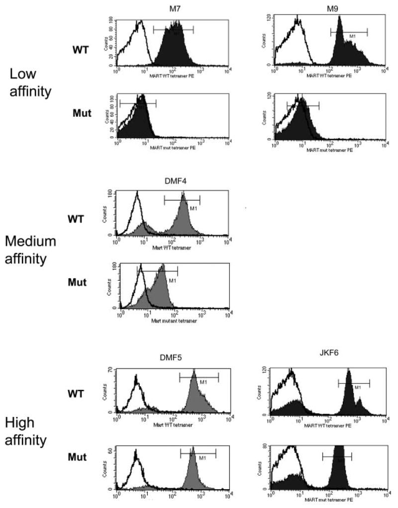
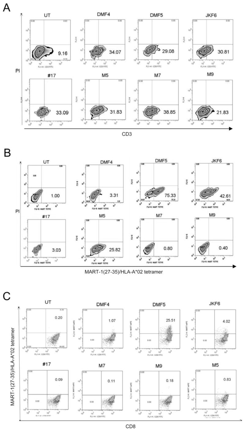
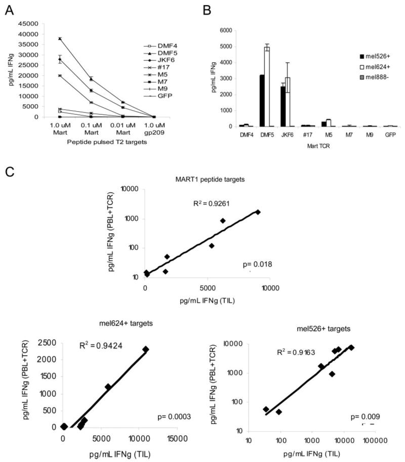
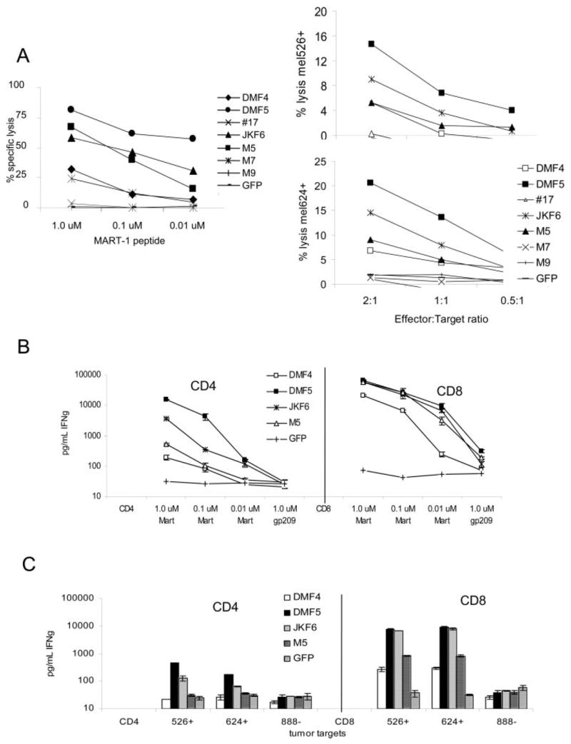
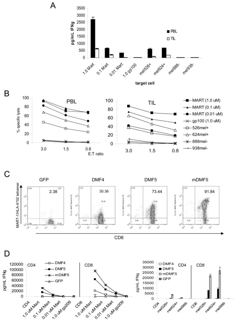
Cell-based antitumor immunity is driven by CD8(+) cytotoxic T cells bearing TCR that recognize specific tumor-associated peptides bound to class I MHC molecules. Of several cellular proteins involved in T cell:target-cell interaction, the TCR determines specificity of binding; however, the relative amount of its contribution to cellular avidity remains unknown. To study the relationship between TCR affinity and cellular avidity, with the intent of identifying optimal TCR for gene therapy, we derived 24 MART-1:27-35 (MART-1) melanoma Ag-reactive tumor-infiltrating lymphocyte (TIL) clones from the tumors of five patients. These MART-1-reactive clones displayed a wide variety of cellular avidities. alpha and beta TCR genes were isolated from these clones, and TCR RNA was electroporated into the same non-MART-1-reactive allogeneic donor PBMC and TIL. TCR recipient cells gained the ability to recognize both MART-1 peptide and MART-1-expressing tumors in vitro, with avidities that closely corresponded to the original TCR clones (p = 0.018-0.0003). Clone DMF5, from a TIL infusion that mediated tumor regression clinically, showed the highest avidity against MART-1 expressing tumors in vitro, both endogenously in the TIL clone, and after RNA electroporation into donor T cells. Thus, we demonstrated that the TCR appeared to be the core determinant of MART-1 Ag-specific cellular avidity in these activated T cells and that nonreactive PBMC or TIL could be made tumor-reactive with a specific and predetermined avidity. We propose that inducing expression of this highly avid TCR in patient PBMC has the potential to induce tumor regression, as an "off-the-shelf" reagent for allogeneic melanoma patient gene therapy.


Conflict of interest statement

Disclosures

The authors have no financial conflict of interest.

Figures

FIGURE 1
FIGURE 1
MART-1-reactive TIL clones derived from several patients display a wide diversity of peptide and MART-1 tumor reactivities. A, IFN-γ production by 24 MART-1 clones derived from five patients, coincubated with titered MART-1 peptide on T2 target cells. B, IFN-γ response of MART-1 clones following coculture with MART-1/HLA-A*0201-positive (mel526+, mel624+) and HLA-A*0201-negative melanoma tumors (mel888, mel938). IFN-γ release was evaluated from 18-h culture supernatant by ELISA. C, Correlation of IFN-γ produced by each clone in response to coculture with T2 cells pulsed with MART-1 peptide or mel624+ MART-1-expressing tumor. D, Correlation of IFN-γ produced by each clone in response to coculture with two different MART-1/HLA-A*0201-expressing tumors. Statistical analysis by ANOVA regression analysis.
FIGURE 2
FIGURE 2
Different MART-1-reactive TIL clones vary in their ability to bind MART-1/HLA-A*0201 tetramer with CD8 independence. Each MART-1-reactive TIL clone was stained with MART-1:26–35(27L)/HLA-A*0201 (MART-1) unmodified tetramer (WT), or MART-1 tetramer with a HLA H chain double mutation abrogating CD8 binding to the HLA-A*0201 molecule (Mut). Representative graphs of each type of TCR binding to tetramer are shown: low-affinity clones were unable to bind tetramer in absence of CD8 coreceptor binding, medium-affinity clones bound CD8-independent tetramer at low levels, and high-affinity clones bound tetramer at high levels in absence of CD8 coreceptor binding. In each graph the isotype control is shown (white) as well as MART-1 tetramer binding (shaded).
FIGURE 3
FIGURE 3
RNA electroporation of TCR derived from MART-1 clones into Jurkat and donor CD8+ PBMC resulted in appropriate αβ TCR translation and surface expression. Percentage of live cells staining positive is indicated. A and B, Two micrograms per 1 × 10⁶ cells in vitro transcribed RNA from TCR α- and β-chains of each MART-1 TIL clone was electroporated into TCR-deficient, CD8-negative Jurkat T3.5 cells that were then stained for CD3 cell surface expression (A) or MART-1: 26–35(27L)/HLA-A*0201 tetramer (B), and evaluated by flow cytometry. C, Flow cytometric evaluation of CD8+ donor PBMC electroporated with 2 μg per 1 × 10⁶ cells TCR RNA, costained with CD8 and MART-1:26–35(27L)/HLA-A*0201 tetramer. UT = untreated cells.
FIGURE 4
FIGURE 4
RNA electroporation of MART-1 TCR derived from clones of varying avidities into activated donor CD8+ PBMC resulted in similar Ag-specific functional avidities. A and B, Donor CD8+-enriched PBMC previously activated with OKT-3 Ab were electroporated with 2 μg of RNA from each TCR chain per 1 × 10⁶ cells, and cocultured overnight with MART-1 peptide pulsed T2 target cells (A) or MART-1/HLA-A*0201-expressing (mel526+, mel624+) or nonexpressing (mel888) melanoma tumors (B). IFN-γ production was measured in supernatants by ELISA. Values shown are the average of duplicate samples, ±SEM. C, Correlation of IFN-γ produced by native TIL clones and donor CD8+ PBMC electroporated with matched TCR RNA. Responses to cocultures with T2 cells pulsed with MART-1 peptide, mel526+, and mel624+ MART-1-expressing melanoma tumors are shown. The p values were obtained by ANOVA regression analysis.
FIGURE 5
FIGURE 5
TCR transfer of a high-avidity TCR in vitro can confer antitumor CTL activity and IFN-γ production in a CD8-independent fashion. A, OKT-3 stimulated CD8+ PBMC RNA-electroporated with different avidity MART-1 TCR were evaluated for their ability to lyse 51Cr-labeled target cells in a 4-h assay. CTL were coincubated with (left) MART-1 peptide-pulsed T2 target cells at 2:1 E:T ratio, or (right) at varying E:T ratios against MART-1 expressing tumor cells. B and C, OKT-3-stimulated donor PBMC were separated into CD4+ and CD8+ populations by positive selection using magnetic beads and each of the four highest avidity MART-1 TCR were expressed in each population by RNA electroporation (2 μg/1 × 106 cells). Functional antitumor response was evaluated by coculture with either MART-1-pulsed T2 target cells (B) or MART-1-expressing tumors (C). IFN-γ production was measured in supernatants by ELISA. Values shown are the average of duplicate samples, ±SEM.
FIGURE 6
FIGURE 6
Nonreactive TIL can also be rendered Ag reactive by TCR transfer of the high-avidity TCR DMF5 in vitro, and this TCR can be altered to generate even higher cellular avidity. A and B, Non-tumor-reactive PBL and TIL from the same patient were stimulated using OKT-3 and irradiated feeder cells in the presence of high-dose IL-2 for 3 days before CD8+ T cell enrichment using magnetic beads. These CD8+ T cells were then electroporated with 2 μg/1 × 106 cells TCR RNA from high-avidity DMF5, and cocultured with MART-1 peptide-pulsed T2 target cells or MART-1/HLA-A*0201-expressing melanoma tumors (mel526+, mel624+) or non-HLA-A*0201-expressing tumors (mel888, mel938). A, IFN-γ production was measured in supernatants by ELISA. Values shown are the average of duplicate samples, ±SEM. B, TIL and PBL electroporated with DMF5 TCR RNA as in A above, were cocultured against MART-1 peptide pulsed T2 cells or tumors in a 4-h 51Cr release assay. Results are representative of four patient samples tested. C and D, The DMF5 TCR was altered by replacing the constant region with murine TCR constant regions (mDMF5) and compared with the naturally occurring DMF5 and DMF4 TCRs by RNA electroporation in activated CD4+ or CD8+ PBMC. C, CD8 and MART-1/HLA-A*0201 tetramer staining of OKT-3-stimulated, TCR RNA-electroporated CD8+ PBMC. Percentage of live lymphocytes staining double positive is indicated. D, IFN-γ production by 18-h coculture of CD4+ and CD8+ TCR RNA electroporated PBMC with MART-1 peptide pulsed T2 targets (left) or tumor target cells (right). Values shown are the average of duplicate samples, ±SEM.

References

    1. Rosenberg SA, Spiess P, Lafreniere R. A new approach to the adoptive immunotherapy of cancer with tumor-infiltrating lymphocytes. Science. 1986;233:1318–1321. - PubMed
    1. Boon T, Coulie PG, Eynde BJ, Bruggen PV. Human T cell responses against melanoma. Annu Rev Immunol. 2006;24:175–208. - PubMed
    1. Rosenberg SA. Progress in human tumour immunology and immunotherapy. Nature. 2001;411:380–384. - PubMed
    1. Dudley ME, Wunderlich JR, Robbins PF, Yang JC, Hwu P, Schwartzentruber DJ, Topalian SL, Sherry R, Restifo NP, Hubicki AM, et al. Cancer regression and autoimmunity in patients after clonal repopulation with antitumor lymphocytes. Science. 2002;298:850–854. - PMC - PubMed
    1. Dudley ME, Wunderlich JR, Yang JC, Sherry RM, Topalian SL, Restifo NP, Royal RE, Kammula U, White DE, Mavroukakis SA, et al. Adoptive cell transfer therapy following non-myeloablative but lymphodepleting chemotherapy for the treatment of patients with refractory metastatic melanoma. J Clin Oncol. 2005;23:2346–2357. - PMC - PubMed

Publication types