. 2006 Oct 31;103(44):16224-9.
doi: 10.1073/pnas.0607640103. Epub 2006 Oct 20.

Yeast mitochondrial ADP/ATP carriers are monomeric in detergents

Affiliations

Yeast mitochondrial ADP/ATP carriers are monomeric in detergents

Lisa Bamber et al. Proc Natl Acad Sci U S A. .

Abstract

Mitochondrial carriers are believed widely to be homodimers both in the inner membrane of the organelle and in detergents. The dimensions and molecular masses of the detergent and protein-detergent micelles were measured for yeast ADP/ATP carriers in a range of different detergents. The radius of the carrier at the midpoint of the membrane, its average radius, its Stokes' radius, its molecular mass, and its excluded volume were determined. These parameters are consistent with the known structural model of the bovine ADP/ATP carrier and they demonstrate that the yeast mitochondrial ADP/ATP carriers are monomeric in detergents. Therefore, models of substrate transport have to be considered in which the carrier operates as a monomer rather than as a dimer.


Conflict of interest statement

The authors declare no conflict of interest.

Figures

Fig. 1.
Fig. 1.
Determination of the molecular masses of AAC2 and AAC3 in alkyl-maltosides by size-exclusion chromatography. (A) The traces for AAC2 in 12M in the absence of CATR and in the presence of CATR are shown in blue and red, respectively. (B) The traces for 8M–13M are shown in red, orange, yellow, green, blue, and purple, respectively. The elution position of standards are indicated by triangles; they are: peaks 1, ferritin (molecular mass 440 kDa, Stokes' radius 61.0 Å); 2, catalase (232 kDa, 52.2 Å); 3, aldolase (158 kDa, 48.1 Å); 4, albumin (67 kDa, 35.5 Å); 5, ovalbumin (43 kDa, 30.5 Å); 6, chymotrypsinogen A (25 kDa, 20.9 Å).
Fig. 2.
Fig. 2.
Relationships between micellar dimensions, molecular masses and the number of detergent molecules associated with AAC3. Stokes' radius (A) and molecular mass (B) of the detergent-protein micelle against the number of detergent molecules associated with AAC3 are shown. The association numbers were based on the weight ratio (filled symbols and dotted line) or on the molecular mass of the protein detergent micelle (open symbols and continuous line). Molecular mass (C) and Stokes' radius (D) of the detergent micelle against the values of the protein–detergent micelle (Table 1) are shown. Symbols for 8M–13M are triangle, inverted triangle, square, diamond, hexagon, and circle, respectively. The standard deviation in the determination of the molecular mass and the Stokes' radius were <3% and 1% of the mean, respectively (n = 2, 3, or 4).
Fig. 3.
Fig. 3.
Sedimentation equilibrium analysis of AAC2 in 12M. (A) Superimposed plots of weight average molecular mass (w,app) against concentration for each cell (datum points), with a line showing the plot for a nonideal monomer (M1 = 32 kDa, B = 2,000 M−1). (B) Plots of residuals between original data and calculated values (for the model above) against radius for each individual cell.
Fig. 4.
Fig. 4.
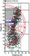
Comparison of dimensions of AAC3 in detergent with the structure of the bovine ADP/ATP carrier AAC1. The distances to the pseudo three-fold axis of symmetry of all Cα atoms (red circles) and other atoms (empty circles) are plotted against the vertical height of the atom. The coordinates of the atoms are taken from the structure of the bovine ADP/ATP carrier AAC1 (Protein Data Bank entry 1OKC) (5). The gray area indicates the void of the cavity, and the red dot indicates the centroid of AAC1. The approximate position of the borders of the membrane (continuous line) and its midpoint (dashed line) are indicated in purple. The determined values for the Stokes' radius of the AAC3 are shown in red. The average radius Rav in 9M and 13M are shown in green and the radii at the midpoint of the membrane as determined for 9M and for the alkyl-maltosides by extrapolation are shown in blue.
Fig. 5.
Fig. 5.
Structure of the bovine AAC1 and dimensions of the detergent micelles and the AAC3-detergent micelles for alkyl-maltosides and digitonin. (A–C) Detergent (A) and AAC3-detergent (B and C) micelles approximated by rounded discs or oblate spheroids, respectively. The rounded disk is likely to be an overestimation of the volume, because it assumes that the radius of the protein does not change with height, whereas the oblate spheroid is likely to be underestimation, because the cross-section is not semicircular. The boundaries of the micelles for 8M-13M are in red, orange, yellow, green, blue, and purple, respectively, and the boundary for digitonin is in brown. The surface representation of AAC1 (Protein Data Bank entry 1OKC) (5) has been rendered in Pymol. Hydrophobic amino acid residues (leucine, valine, isoleucine, tryptophan, methionine, cysteine, phenylalanine, and alanine) are orange. Molecules of 8M, 10M, and digitonin are shown in red, yellow, and black carbon spheres for size comparisons. For digitonin and 8M, the excluded volume is represented by a cylinder with radius Rav, shown in projection by a dotted and solid black line, respectively (Table 3 and Eq. 12). The difference in excluded volume between successive micelles in the alkyl maltoside series is represented by two cylinders at the interface above and below, each with height ΔRdm and calculated average radius according to Eq. 13. All of the volumes that were not excluded by the protein in the protein–detergent micelle are colored blue and they most likely represent water cavities. (Scale bar, 10 Å.)

References

    1. Palmieri F. Pflugers Arch. 2004;447:689–709. - PubMed
    1. Kunji ER. FEBS Lett. 2004;564:239–244. - PubMed
    1. Saraste M, Walker JE. FEBS Lett. 1982;144:250–254. - PubMed
    1. Kunji ER, Harding M. J Biol Chem. 2003;278:36985–36988. - PubMed
    1. Pebay-Peyroula E, Dahout-Gonzalez C, Kahn R, Trezeguet V, Lauquin GJ, Brandolin G. Nature. 2003;426:39–44. - PubMed

Publication types

LinkOut - more resources