. 2006 Dec;5(12):2092-103.
doi: 10.1128/EC.00288-06. Epub 2006 Oct 20.

Nucleocytoplasmic shuttling of the Rpb4p and Rpb7p subunits of Saccharomyces cerevisiae RNA polymerase II by two pathways

Affiliations

Nucleocytoplasmic shuttling of the Rpb4p and Rpb7p subunits of Saccharomyces cerevisiae RNA polymerase II by two pathways

Michael Selitrennik et al. Eukaryot Cell. 2006 Dec.

Abstract

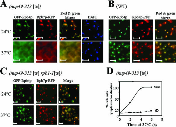
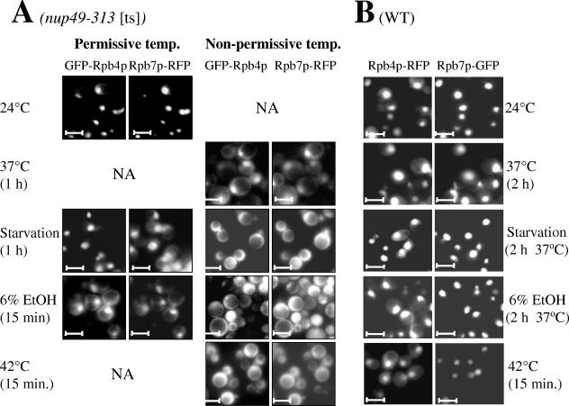
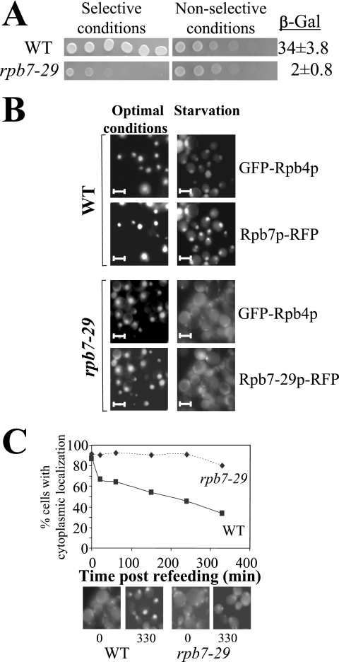
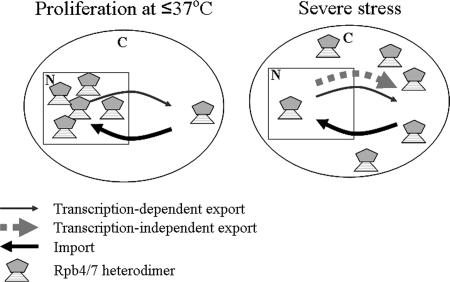
Rpb4p and Rpb7p are subunits of the RNA polymerase II of Saccharomyces cerevisiae that form a dissociable heterodimeric complex. Whereas the only reported function of Rpb7p is related to transcription, Rpb4p has been found to also act in mRNA export and in the major mRNA decay pathway that operates in the cytoplasm, thus raising the possibility that Rpb4p links between the nuclear and cytoplasmic processes. Here we show that both Rpb4p and Rpb7p shuttle between the nucleus and the cytoplasm. Shuttling kinetics of the two proteins are similar as long as their interaction is possible, suggesting that they shuttle as a heterodimer. Under normal conditions, shuttling of Rpb4p and Rpb7p depends on ongoing transcription. However, during severe stresses of heat shock, ethanol, and starvation, the two proteins shuttle via a transcription-independent pathway. Thus, Rpb4p and Rpb7p shuttle via two pathways, depending on environmental conditions.


Figures

FIG. 1.
FIG. 1.
Rpb7p-GFP localization is responsive to the environment. (A) Rpb7p is exported to the cytoplasm in response to various stress conditions. Cells carrying RPB7-GFP in place of RPB7 (yMC286) were allowed to proliferate for at least eight generations under optimal conditions in rich medium (YPD) at either 24°C (HS experiment) or 30°C (other experiments) until the mid-logarithmic phase (≤5 × 10⁶ cells/ml). Cells were then treated with 50 μg/ml cycloheximide except for the starvation culture and were challenged with stress as indicated. The nonstressed samples were also treated with 50 μg/ml cycloheximide and were further incubated in parallel with the stress-treated cultures. “Starvation” denotes synthetic medium lacking sugar and amino acids. Rpb7p-GFP localization was examined by fluorescent microscopy 3 h after HS or 12 h after addition of EtOH or after 3 days of starvation. The middle panels of the HS experiment show the position of the nuclei stained by DAPI (4′,6′-diamidino-2-phenylindole), and the bottom panels show bright-field (BF) images of the cells; the results shown demonstrate that Rpb7p localization during optimal proliferation is nuclear whereas after HS it is cytoplasmic. (B) Cytoplasmic Rpb7p-GFP in starved cells was reimported to the nucleus after cells were refed with glucose. Starved cells were refed with 2% glucose and divided into two cultures. One culture (designated CHX) was treated with 50 μg/ml cycloheximide, and the cells were examined microscopically 90 min after addition of glucose. In the absence of the drug, the kinetics of Rpb7p-GFP relocation was similar to that observed in its presence (results not shown). Note that in response to starvation (see panels A and B), Rpb7p-GFP was localized in discrete cytoplasmic foci that are smaller than nuclei. Some are denoted by arrows. These foci probably represent cytoplasmic P bodies that also accommodate Rpb4p (17). The bars represent 5μ.
FIG. 2.
FIG. 2.
GFP-Rpb4p and Rpb7p-RFP shuttle by a transcription-dependent mechanism. (A) Shuttling of both proteins was demonstrated using nup49-313(ts) mutant cells that are defective in protein import at elevated temperatures. Cells expressing both GFP-Rpb4p and Rpb7p-RFP (yMS3) were allowed to proliferate for at least eight generations under optimal conditions in selective media at 24°C. Both fluorescent proteins were then localized in the nucleus (results not shown). Cycloheximide (50 μg/ml) was then added, and the culture was divided into two samples. One was incubated for an additional 5 h at 24°C and the other for 5 h at 37°C. Images were then taken using green (GFP-Rpb4p), red (Rpb7p-RFP), or blue (to detect nuclei stained by DAPI) filters, as indicated. (B) Images of WT cells expressing Rpb4p-RFP and Rpb7p-GFP (yMS8) at 24°C or 5 h after incubation at 37°C in the presence of 50 μg/ml cycloheximide. (C) Export is blocked when transcription is arrested. Images of nup49-313(ts) rpb1-1(ts) cells expressing both GFP-Rpb4p and Rpb7p-RFP (yMS4) at 24°C or 5 h after incubation at 37°C are shown. Cycloheximide was added as described for panel A. (D) Export kinetics of Rpb7p-RFP after a shift from 24°C to 37°C in the absence (Cont.) or presence (Φ) of the transcription inhibitor 1,10-phenanthroline (100 μg/ml). Cycloheximide was added as described for panel A. To monitor export kinetics, samples of each culture were examined microscopically at the indicated time points and photographs of random fields were taken using both the green and red channels. More than 200 cells were classified into those exhibiting nuclear or whole-cell (i.e., cytoplasmic) localization of the respective fluorescent protein. The proportion of cells exhibiting cytoplasmic localization was plotted as a function of time. The merged images in panels A and C (created using Adobe PhotoShop) demonstrate that the distributions of the two fluorescent proteins were similar. The bars represent 5μ.
FIG. 3.
FIG. 3.
Export of GFP-Rpb4p and Rpb7p-RFP during severe stresses is independent of transcription. (A) Images of nup49-313(ts) rpb1-1(ts) cells expressing both GFP-Rpb4p and Rpb7p-RFP (yMS4) in the mid-logarithmic phase at 24°C (left panels) or after 2 h under the indicated stress conditions in the presence of 50 μg/ml cycloheximide. The nonstressed sample was equally treated with cycloheximide and further incubated in parallel with the stress-treated cultures. The starved and EtOH-treated cultures were incubated at 37°C to inactivate transcription. The bars represent 5μ. (B) Export kinetics, determined as described for Fig. 2D, of Rpb7p-RFP after a shift from 24°C to 42°C in the absence (Cont.) or presence (Φ) of the transcription inhibitor 1,10-phenanthroline (100 μg/ml).
FIG. 4.
FIG. 4.
Rpb4p and Rpb7p shuttle during severe stress conditions. (A) Relocation in nup49-313 cells is faster at the nonpermissive temperature. nup49-313(ts) cells expressing both GFP-Rpb4p and Rpb7p-RFP (yMS3) were allowed to proliferate for at least eight generations under optimal conditions in selective media at 24°C. Cells were then challenged with the indicated stress at 24°C (Permissive temp.) (left panels) or at 37°C, or 42°C when indicated (Non-permissive temp.) (right panels). Cycloheximide (50 μg/ml) was added to all samples as described for Fig. 2A. (B) Little relocation occurs in WT cells. Images of WT cells expressing both GFP-Rpb4p and Rpb7p-RFP (yMS8) similarly challenged with the indicated conditions in the presence of cycloheximide are shown. NA, not applicable. The bars represent 5μ.
FIG. 5.
FIG. 5.
In response to various stresses, Rpb4p and Rpb7p are exported to the cytoplasm at similar kinetics. (A to C) yMS4 (A), yMS18 (B), and yMS8 (C) cells expressing the indicated fluorescence-tagged proteins were allowed to proliferate for at least eight generations under optimal conditions in selective media at either 24°C (A) or 30°C (B, C, and D) until the mid-logarithmic phase (5 × 10⁶ cells/ml). Cultures were then challenged under the indicated stress condition as described for Fig. 1. Export kinetics experiments were performed as described for Fig. 2D. (D) Import kinetics of GFP-Rpb4p and Rpb7p-RFP are comparable. yRL82 cells were starved until both fluorescent proteins were localized in the cytoplasm in 80% or 90% of the cells. Cells were then collected by centrifugation and resuspended in synthetic complete medium (containing glucose and amino acids) supplemented with 50 μg/ml cycloheximide (similar results were obtained when cycloheximide was not added). Import kinetics results were determined as described for panels A to C.
FIG. 6.
FIG. 6.
Transport of Rpb7p-GFP during severe stresses is affected by RPB4. (A) Stress-induced relocation of Rpb7p-GFP is accelerated in the absence of RPB4. Images of WT cells carrying RPB7-GFP in place of RPB7 (yMC286) and isogenic rpb4Δ-derivative cells (yMC310) that were challenged with various stresses for relatively short periods of time are shown. The kinds of stresses and their durations are indicated on the right side of each panel. Starvation conditions were simulated by use of synthetic medium lacking sugar and amino acids. Note that after a brief period of stress, relatively little export was detected in the WT cells (left panels) whereas export in the mutant was more advanced (right panels). (B) Efficient import of Rpb7p-GFP is dependent on Rpb4p. The strains described for panel A were incubated in starvation medium for 72 h to permit relocation of Rpb7p-GFP in 77% of WT cells and 100% of the mutant cells. Cells were then collected by centrifugation and resuspended in synthetic complete medium (containing glucose and amino acids) supplemented with 50 μg/ml cycloheximide. Import kinetics results were determined as described for Fig. 2. Note that in response to starvation or HS, Rpb7p-GFP can be localized in discrete cytoplasmic foci that are smaller than nuclei. We suspect that these foci represent cytoplasmic P bodies (17).
FIG. 7.
FIG. 7.
Transport of GFP-Rpb4p during starvation is affected by RPB7. (A) Two-hybrid interaction between Rpb7p-DBD and Rpb4-AD (upper row) and between Rpb7-29p-DBD and Rpb4p-AD (lower row). Cells were spotted in threefold serial dilutions starting with 106 cells/spot on an indicator plate (Selective conditions) as described in Materials and Methods. Growth on this plate indicates interaction (38). To demonstrate spotting of equal amounts of cells, cells were spotted on a nonselective plate that allowed growth of cells that carry both the bait and prey plasmids independently of the two two-hybrid interactions (using medium lacking only leucine and tryptophane). Beta-galactosidase (β-Gal) values, quantitative means used to determine interactions (38), are indicated at the right. They reflect the average values determined for three independent experiments. Background values obtained with cells expressing only the bait plasmid were subtracted. (B) Starvation-induced relocation of GFP-Rpb4p and Rpb7p-RFP is faster in rpb7-29 cells than in WT cells. Images of WT cells carrying RPB7-RFP in place of RPB7 and GFP-RPB4 (yRL82) or cells carrying a rpb7-29-RFP derivative thereof (yRL81) that were challenged with starvation (using a synthetic medium lacking sugar and amino acids) at 37°C for a relatively short period of time (45 min) are shown. The same field was photographed using either the green (for detection of GFP-Rpb4p) or red (for detection of Rpb7p-RFP) channel, as indicated. Note that after a short period of starvation, relatively little export was detected in the WT cells (upper right panels) whereas export of both fluorescent proteins in the mutant was more advanced (lower right panels). (C) Import of GFP-Rpb4p is impaired in rpb7-29 mutant cells. WT cells (yRL82) or rpb7-29 mutant cells (yRL81) expressing GFP-RPB4 were starved until GFP-Rpb4p was localized in the cytoplasm in ∼90% of the cells. Cells were then collected by centrifugation and resuspended in synthetic complete medium (containing glucose and amino acids) supplemented with 50 μg/ml cycloheximide (similar results were obtained when cycloheximide was not added). Import kinetics results were determined as described for Fig. 5D. Examples of photographs of cells taken at the indicated time points after refeeding are shown below the graph.
FIG. 8.
FIG. 8.
Model of the proposed shuttling pathways of the Rpb4-Rbp7 heterodimer. Rpb4p and Rpb7p shuttle as a heterodimer. Under conditions that support proliferation, export of the Rpb4-Rbp7 heterodimer is dependent on transcription. Import of the heterodimer (thick arrow) is faster than its export (thin arrow), resulting in mainly nuclear localization. In response to some severe stress conditions, a relatively robust transcription-independent mechanism is induced (thick dashed arrow) in addition to a relatively weak transcription-dependent mechanism (thin arrow). Export is then faster than import, and the general direction of the net flow is from the nucleus to the cytoplasm. Because the Rpb4-Rbp7 heterodimer must be located in the nucleus to enable transcription during stress (5), this flux of the Rpb4-Rbp7 heterodimer from the nucleus to the cytoplasm must be leveled off at some point during prolonge incubation under these stress conditions and balanced by the retrograde flux. While export of Rpb4p or Rpb7p does not seem to be inhibited by disruption of the heterodimer integrity, the import pathway is very sensitive to this integrity. Thus, import of Rpb4p or Rpb7p is severely compromised when the heterodimer is disrupted either by deleting RPB4 or by mutating RPB7. Import is also blocked by inactivating Nup49p. In all these cases the net flow of these molecules from the nucleus to the cytoplasm is faster than normal. N denotes the nucleus; C denotes the cytoplasm.

References

    1. Aguilera, A. 2005. Cotranscriptional mRNP assembly: from the DNA to the nuclear pore. Curr. Opin. Cell Biol. 17:242-250. - PubMed
    1. Armache, K. J., H. Kettenberger, and P. Cramer. 2003. Architecture of initiation-competent 12-subunit RNA polymerase II. Proc. Natl. Acad. Sci. USA 100:6964-6968. - PMC - PubMed
    1. Bushnell, D. A., and R. D. Kornberg. 2003. Complete, 12-subunit RNA polymerase II at 4.1-A resolution: implications for the initiation of transcription. Proc. Natl. Acad. Sci. USA 100:6969-6973. - PMC - PubMed
    1. Choder, M. 1993. A growth rate-limiting process in the last growth phase of the yeast life cycle involves RPB4, a subunit of RNA polymerase II. J. Bacteriol. 175:6358-6363. - PMC - PubMed
    1. Choder, M. 2004. Rpb4 and Rpb7: subunits of RNA polymerase II and beyond. Trends Biochem. Sci. 29:674-681. - PubMed

Publication types

MeSH terms