. 2007 Jan;189(2):369-76.
doi: 10.1128/JB.01373-06. Epub 2006 Oct 20.

Coordinate expression of the acetyl coenzyme A carboxylase genes, accB and accC, is necessary for normal regulation of biotin synthesis in Escherichia coli

Affiliations

Coordinate expression of the acetyl coenzyme A carboxylase genes, accB and accC, is necessary for normal regulation of biotin synthesis in Escherichia coli

Ahmed M Abdel-Hamid et al. J Bacteriol. 2007 Jan.

Abstract

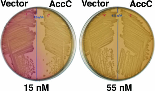
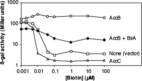
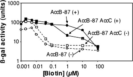
Transcription of the biotin (bio) biosynthetic operon of Escherichia coli is negatively regulated by the BirA protein, an atypical repressor protein in that it is also an enzyme. The BirA-catalyzed reaction involves the covalent attachment of biotin to AccB, a subunit of acetyl coenzyme (acetyl-CoA) carboxylase. The two functions of BirA allow regulation of the bio operon to respond to the intracellular concentrations of both biotin and unbiotinylated AccB. We report here that bio operon expression is down-regulated by overproduction of AccC, another acetyl-CoA carboxylase subunit known to form a complex with AccB. This down-regulation is eliminated when AccB and AccC are coordinately overexpressed, but only when the AccB partner is competent to bind AccC. Under AccC overexpression conditions AccB is underbiotinylated. These findings can be explained by a model in which excess AccC sequesters AccB in a complex that is a poor substrate for biotinylation. The observed disruption of biotin synthesis and attachment provides an excellent rationale for the observation that in the vast majority of sequenced bacterial genomes AccB and AccC are encoded in a two-gene operon.


Figures

FIG. 1.
FIG. 1.
Biotin regulatory system of E. coli. BirA is represented by green ovals, biotin is represented by black circles, the AMP moiety is represented by red pentagons, AccB is represented by dark blue ovals, and AccC is represented by light blue crescents. The arrows indicate transcription from the leftward and rightward bio promoters. (A to C) BirA switches from the biotin ligation function to the repressor function in response to the intracellular biotin requirement, which is indicated by the level of unbiotinylated AccB. If unbiotinylated AccB levels are high, the protein functions as a biotin ligase. Once the unbiotinylated AccB has been converted to the biotinylated form, the biotinoyl-5′-AMP is no longer consumed and remains bound to BirA. The liganded form of BirA accumulates to levels sufficiently high that the bio operator is fully occupied, resulting in transcriptional repression of the biotin biosynthetic genes. (D) Overproduction of AccC ties up unbiotinylated AccB in a complex that is a poor biotinylation substrate. Therefore, high levels of the liganded form of BirA accumulate, resulting in repression of bio operon transcription.
FIG. 2.
FIG. 2.
BirA and acetyl-CoA carboxylase reactions. (A) BirA reaction, which is the general reaction of biotin protein ligases (9). (B) Acetyl-CoA carboxylase reaction. The functional enzyme is thought to have the composition 2AccA:4AccB:2AccC:2AccD, with the AccB-AccC and AccA-AccD complexes sufficiently stable for isolation in vitro (18).
FIG. 3.
FIG. 3.
Repression of bio operon transcription upon overproduction of AccC. The plates are MacConkey agar plates containing 100 μM IPTG, and the biotin concentrations are indicated below the plates. The red color is due to depression of the bioBFCD promoter that drives expression of the lacZ and lacY genes (4). The host was strain CY1740, and the accC and vector plasmids were pCY701 and pKK223-3, respectively. Essentially identical results were obtained with strains BM2661 and CY486.
FIG. 4.
FIG. 4.
Expression with various biotin concentrations of the chromosomal φ(bioFC-lacZ)501 fusion of derivatives of strain CY1740 carrying a plasmid encoding AccB (○) or AccC (▵) or the vector pKK223-3 (□). A derivative carrying two compatible plasmids, one encoding AccB and the other encoding BirA (•), was also assayed. The accC, accB, and birA plasmids were pCY701, pCY708, and pCY216, respectively. Note that both axes have log scales. The experiment was repeated twice in its entirety, and in addition, the vector and AccC overproduction cultures were compared two more times. The results were essentially identical to those shown. β-gal, β-galactosidase.
FIG. 5.
FIG. 5.
Expression with various biotin concentrations of the chromosomal φ(bioFC-lacZ)501 fusion of derivatives of strain CY1740 carrying a plasmid encoding AccBC (▪ and □) or AccC (• and ○). The solid symbols and plus signs indicate induction by addition of 0.1 mM IPTG, whereas the open symbols and minus signs indicate uninduced cultures. The accC and accBC plasmids were pCY701 and pCY705, respectively. The experiment was repeated twice in its entirety, and in addition, the AccBC and AccC overproduction cultures were compared two more times. The results were essentially identical to those shown. β-gal, β-galactosidase.
FIG. 6.
FIG. 6.
Expression with various biotin concentrations of the chromosomal φ(bioFC-lacZ)501 fusion of derivatives of strain CY1740 carrying plasmid pAH1 encoding the carboxyl 87 residues of AccB (AccB-87) (• and ○) or plasmid pAH2 encoding AccB-87 plus AccC (▪ and □). The solid symbols and plus signs indicate induction by addition of 0.1 mM IPTG, whereas the open symbols and minus signs indicate uninduced cultures. The experiment was repeated twice, and the results were essentially identical to those shown. β-gal, β-galactosidase.
FIG. 7.
FIG. 7.
Expression of the chromosomal φ(bioFC-lacZ)501 fusion of derivatives of strain CY1740 carrying plasmids encoding AccC plus mutant AccB proteins. The AccB K122R protein (•), mutant AccB K122E protein (□), and mutant AccB K122M protein (▴) were encoded by plasmids pAH7, pAH13, and pAH14, respectively. The experiment was repeated twice, and the results were essentially identical to those shown. β-gal, β-galactosidase.

References

    1. Alberts, A. W., A. M. Nervi, and P. R. Vagelos. 1969. Acetyl CoA carboxylase. II. Demonstration of biotin-protein and biotin carboxylase subunits. Proc. Natl. Acad. Sci. USA 63:1319-1326. - PMC - PubMed
    1. Barker, D. F., and A. M. Campbell. 1981. The birA gene of Escherichia coli encodes a biotin holoenzyme synthetase. J. Mol. Biol. 146:451-467. - PubMed
    1. Barker, D. F., and A. M. Campbell. 1981. Genetic and biochemical characterization of the birA gene and its product: evidence for a direct role of biotin holoenzyme synthetase in repression of the biotin operon in Escherichia coli. J. Mol. Biol. 146:469-492. - PubMed
    1. Barker, D. F., and A. M. Campbell. 1980. Use of bio-lac fusion strains to study regulation of biotin biosynthesis in Escherichia coli. J. Bacteriol. 143:789-800. - PMC - PubMed
    1. Beckett, D. 2005. The Escherichia coli biotin regulatory system: a transcriptional switch. J. Nutr. Biochem. 16:411-415. - PubMed

Publication types

MeSH terms

LinkOut - more resources