. 2006;2006(4):26818.
doi: 10.1155/JBB/2006/26818.

Intronic microRNA (miRNA)

Affiliations

Intronic microRNA (miRNA)

Shi-Lung Lin et al. J Biomed Biotechnol. 2006.

Abstract

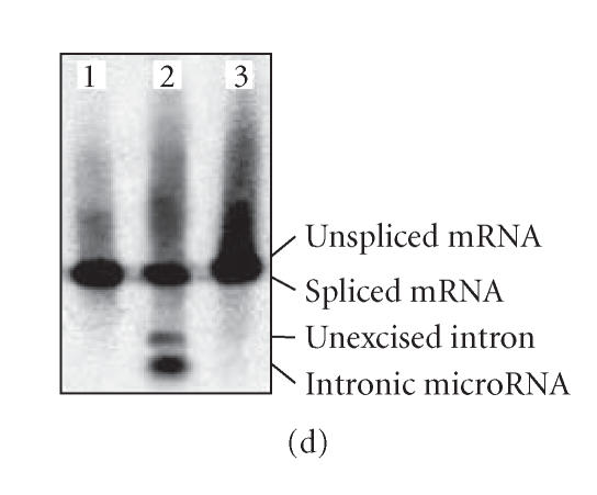
Nearly 97% of the human genome is composed of noncoding DNA, which varies from one species to another. Changes in these sequences often manifest themselves in clinical and circumstantial malfunction. Numerous genes in these non-protein-coding regions encode microRNAs, which are responsible for RNA-mediated gene silencing through RNA interference (RNAi)-like pathways. MicroRNAs (miRNAs), small single-stranded regulatory RNAs capable of interfering with intracellular messenger RNAs (mRNAs) with complete or partial complementarity, are useful for the design of new therapies against cancer polymorphisms and viral mutations. Currently, many varieties of miRNA are widely reported in plants, animals, and even microbes. Intron-derived microRNA (Id-miRNA) is a new class of miRNA derived from the processing of gene introns. The intronic miRNA requires type-II RNA polymerases (Pol-II) and spliceosomal components for their biogenesis. Several kinds of Id-miRNA have been identified in C elegans, mouse, and human cells; however, neither function nor application has been reported. Here, we show for the first time that intron-derived miRNAs are able to induce RNA interference in not only human and mouse cells, but in also zebrafish, chicken embryos, and adult mice, demonstrating the evolutionary preservation of intron-mediated gene silencing via functional miRNA in cell and in vivo. These findings suggest an intracellular miRNA-mediated gene regulatory system, fine-tuning the degradation of protein-coding messenger RNAs.


Figures

Figure 1
Figure 1
Comparison of biogenesis and RNAi mechanisms among siRNA, intergenic (exonic) miRNA, and intronic miRNA. siRNA is likely formed by two perfectly complementary RNAs transcribed from two different promoters (remaining to be determined) and further processing into 19–22 bp duplexes by the RNase III-familial endonuclease, Dicer. The biogenesis of intergenic miRNAs, for example, lin-4 and let-7, involves a long transcript precursor (pri-miRNA), which is probably generated by Pol-II or Pol-III RNA promoters, while intronic miRNAs are transcribed by the Pol-II promoters of its encoded genes and coexpressed in the intron regions of the gene transcripts (pre-mRNA). After RNA splicing and further processing, the spliced intron may function as a pri-miRNA for intronic miRNA generation. In the nucleus, the pri-miRNA is excised by Drosha RNase to form a hairpin-like pre-miRNA template and then exported to the cytoplasm for further processing by Dicer* to form mature miRNA. The Dicers for siRNA and miRNA pathways are different. All three small regulatory RNAs are finally incorporated into an RNA-induced silencing complex (RISC), which contains either the strand of siRNA or the single-strand of miRNA. The action of miRNA is considered to be more specific and less adverse than that of siRNA because only one strand is involved. siRNA primarily triggers mRNA degradation, whereas miRNA can induce either mRNA degradation or suppression of protein synthesis depending on the sequence complementarity to the target gene transcripts.
Figure 2
Figure 2
Biogenesis and function of intronic miRNA. (a) The native intronic miRNA is cotranscribed with a precursor messenger RNA (pre-mRNA) by Pol-II and cleaved out of the pre-mRNA by an RNA splicing machinery, the spliceosome. The spliced intron with hairpin-like secondary structure is further processed into mature miRNA capable of triggering RNAi effects, while the ligated exons become a mature messenger RNA (mRNA) for protein synthesis. (b) We designed an artificial intron containing pre-miRNA, namely SpRNAi, mimicking the biogenesis of the native intronic miRNA. (c) When a designed miR-EGFP(280–302)-stemloop RNA construct was tested in the EGFP-expressing Tg(UAS:gfp) zebrafish, we detected a strong RNAi effect only on the target EGFP (lane 4). No detectable gene-silencing effect was observed in other lanes; from left to right: 1, blank vector control (Ctl); 2, miRNA-stemloop targeting HIV-p24 (mock); 3, miRNA without stemloop (anti); and 5, stemloop-miRNA* complementary to the miR-EGFP(280–302) sequence (miR*). The off-target genes, such as vector RGFP and fish actin, were not affected, indicating the high target specificity of miRNA-mediated gene silencing. (c) Three different miR-EGFP(280–302) expression systems were tested for miRNA biogenesis; from left to right: 1, vector expressing intron-free RGFP, no pre-miRNA insert; 2, vector expressing RGFP with an intronic 5′-miRNA-stemloop-miRNA*-3′ insert; and 3, vector similar to the 2 construct but with a defected 5′-splice site in the intron. In Northern blot analysis probing the miR-EGFP(280–302) sequence, the mature miRNA was released only from the spliced intron resulting from the vector 2 construct in the cell cytoplasm.
Figure 2
Figure 2
Biogenesis and function of intronic miRNA. (a) The native intronic miRNA is cotranscribed with a precursor messenger RNA (pre-mRNA) by Pol-II and cleaved out of the pre-mRNA by an RNA splicing machinery, the spliceosome. The spliced intron with hairpin-like secondary structure is further processed into mature miRNA capable of triggering RNAi effects, while the ligated exons become a mature messenger RNA (mRNA) for protein synthesis. (b) We designed an artificial intron containing pre-miRNA, namely SpRNAi, mimicking the biogenesis of the native intronic miRNA. (c) When a designed miR-EGFP(280–302)-stemloop RNA construct was tested in the EGFP-expressing Tg(UAS:gfp) zebrafish, we detected a strong RNAi effect only on the target EGFP (lane 4). No detectable gene-silencing effect was observed in other lanes; from left to right: 1, blank vector control (Ctl); 2, miRNA-stemloop targeting HIV-p24 (mock); 3, miRNA without stemloop (anti); and 5, stemloop-miRNA* complementary to the miR-EGFP(280–302) sequence (miR*). The off-target genes, such as vector RGFP and fish actin, were not affected, indicating the high target specificity of miRNA-mediated gene silencing. (c) Three different miR-EGFP(280–302) expression systems were tested for miRNA biogenesis; from left to right: 1, vector expressing intron-free RGFP, no pre-miRNA insert; 2, vector expressing RGFP with an intronic 5′-miRNA-stemloop-miRNA*-3′ insert; and 3, vector similar to the 2 construct but with a defected 5′-splice site in the intron. In Northern blot analysis probing the miR-EGFP(280–302) sequence, the mature miRNA was released only from the spliced intron resulting from the vector 2 construct in the cell cytoplasm.
Figure 3
Figure 3
Schematic construct of the artificial SpRNAi intron in a recombinant gene SpRNAi-RGFP for intracellular expression and processing. The components of the Pol-II-mediated SpRNAi system include several consensus nucleotide elements consisting of a 5′-splice site, a branch-point domain (BrP), a poly-pyrimidine tract (PPT), a 3′-splice site, and a pre-miRNA insert located between the 5′-splice site and the BrP domain. The expression of the recombinant gene is under the regulation of either a mammalian Pol-II RNA promoter or a compatible viral promoter for cell-type-specific effectiveness. Mature miRNA molecules are released from the intron by RNA splicing and further Dicer processing.
Figure 4
Figure 4
Intronic miRNA-mediated gene silencing effects in vivo. (a)–(c) Different preferences for RISC assembly were observed following the transfection of 5′-miRNA*-stemloop-miRNA-3′ (1) and 5′-miRNA-stemloop-miRNA*-3′ (2) pre-miRNA structures in zebrafish, respectively. (a) One mature miRNA, namely miR-EGFP(280/302), was detected in the (2)-transfected zebrafish, whereas the (1)-transfection produced another kind of miRNA, miR*-EGFP(301–281), which was partially complementary to the miR-EGFP(280/302). (b) The RNAi effect was only observed with the transfection of the (2) pre-miRNA, showing less EGFP (green) in (2) than in (1), while the miRNA indicator RGFP (red) was equally present in all vector transfections. (c) Western blot analysis of the EGFP protein levels confirmed the specific silencing result of (b). No detectable gene silencing was observed in fish without (Ctl) and with liposome only (Lipo) treatments. The transfection of either a U6-driven siRNA vector (siR) or an empty vector (Vctr) without the designed pre-miRNA insert resulted in no significant gene silencing. (d)–(g) Silencing of endogenous β-catenin and noggin genes in chicken embryos. (d) The pre-miRNA construct and fast green dye mixtures were injected into the ventral side of chicken embryos near the liver primordia below the heart. (e) Northern blot analysis of extracted RNAs from chicken embryonic livers with anti-β-catenin miRNA transfections (lanes 4–6) in comparison with wild types (lanes 1–3) showed a more than 98% silencing effect on β-catenin mRNA expression, while the house-keeping gene, GAPDH, was not affected. (f) Liver formation of the β-catenin knockouts was significantly hindered (upper right 2 panels). Microscopic examination revealed a loose structure of hepatocytes, indicating the loss of cell-cell adhesion due to breaks in adherents junctions formed between β-catenin and cell membrane E-cadherin in early liver development. In severely affected regions, feather growth in the skin close to the injection area was also inhibited (lower right 2 panels). Immunohistochemical staining for β-catenin protein (brown) showed a significant decrease in the feather follicle sheaths. (g) Lower beak development was increased by the mandibular injection of the antinoggin pre-miRNA construct (lowerpanel) in comparison to wild type (upper panel). Right panels showed bone (alizarin red) and cartilage (alcian blue) staining to demonstrate the outgrowth of bone tissues in the lower beak of the noggin knockout. Northern blot analysis (small windows) confirmed a ∼ 60% decrease of noggin mRNA expression in the lower beak area.
Figure 5
Figure 5
Silencing of HIV-1 genome replication using anti-gag/pro/pol miRNA transfection into CD4+ T lymphocytes isolated from the acute and chronic phases of AIDS infections. (a) Northern blot analysis showed about 98% and 70% decreases of HIV genome in the acute and chronic infections after miRNA treatments (lane 4), respectively. No effect was detected in the T cells transfected with miRNA* targeting the same gag/pro/pol region of the viral genome (lane 5). The size of pure HIV-1 provirus was about 9,700 nucleotide bases (lane 1). RNA extracts from normal noninfected CD4+ T lymphocytes were used as a negative control (lane 2), whereas those from HIV-infected T cells were used as a positive control (lane 3). (b) Immunostaining for HIV p24 marker confirmed the results in (a). Since the ex vivo HIV-silenced T lymphocytes were resistant to any further infection by the same strains of HIV, they may be transfused back to the donor patient for eliminating HIV-infected cells.
Figure 5
Figure 5
Silencing of HIV-1 genome replication using anti-gag/pro/pol miRNA transfection into CD4+ T lymphocytes isolated from the acute and chronic phases of AIDS infections. (a) Northern blot analysis showed about 98% and 70% decreases of HIV genome in the acute and chronic infections after miRNA treatments (lane 4), respectively. No effect was detected in the T cells transfected with miRNA* targeting the same gag/pro/pol region of the viral genome (lane 5). The size of pure HIV-1 provirus was about 9,700 nucleotide bases (lane 1). RNA extracts from normal noninfected CD4+ T lymphocytes were used as a negative control (lane 2), whereas those from HIV-infected T cells were used as a positive control (lane 3). (b) Immunostaining for HIV p24 marker confirmed the results in (a). Since the ex vivo HIV-silenced T lymphocytes were resistant to any further infection by the same strains of HIV, they may be transfused back to the donor patient for eliminating HIV-infected cells.

References

    1. Ambros V, Lee RC, Lavanway A, Williams PT, Jewell D. MicroRNAs and other tiny endogenous RNAs in C. elegans. Current Biology. 2003;13(10):807–818. - PubMed
    1. Lin SL, Chang D, Wu D-Y, Ying SY. A novel RNA splicing-mediated gene silencing mechanism potential for genome evolution. Biochemical and Biophysical Research Communications. 2003;310(3):754–760. - PubMed
    1. Rodriguez A, Griffiths-Jones S, Ashurst JL, Bradley A. Identification of mammalian microRNA host genes and transcription units. Genome Research. 2004;14(10 A):1902–1910. - PMC - PubMed
    1. Lin SL, Chuong CM, Ying SY. A novel mRNA-cDNA interference phenomenon for silencing bcl-2 expression in human LNCaP cells. Biochemical and Biophysical Research Communications. 2001;281(3):639–644. - PubMed
    1. Ying SY, Lin SL. Intron-derived microRNAs - fine tuning of gene functions. Gene. 2004;342(1):25–28. - PubMed

LinkOut - more resources