

Effect of death-to-preservation time on donor corneal epithelium
- PMID: 17057804
- PMCID: PMC1447575
Effect of death-to-preservation time on donor corneal epithelium
Abstract
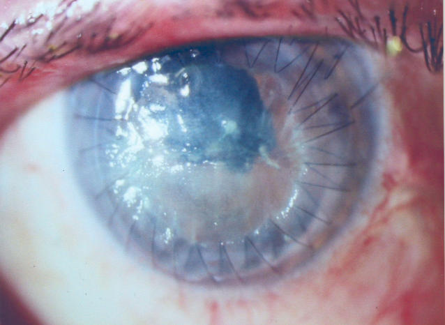
Purpose: Surface disease is one of multiple variables affecting the quality of the postkeratoplasty donor cornea. Trauma to Bowman's layer before and during harvesting can denude the donor epithelium and result in epithelial defects in the donor following penetrating keratoplasty. Eye banks use death-to-preservation (DP) time intervals as long as 18 hours. This study evaluates the effects of higher DP time on the donor epithelium in storage medium and immediately following keratoplasty.
Methods: Eighty-one consecutive corneas were procured by the University of Kentucky Eye Bank, rated by one technician (H.W.), and used by one surgeon (W.S.V.) for elective penetrating keratoplasty. Donor records were retrospectively reviewed for age, DP time, and epithelial condition. All corneas were harvested and evaluated according to Eye Bank Association of America standards. Donor charts were reviewed for DP time and for condition of the epithelium in storage. Recipient charts were reviewed for epithelial defects following keratoplasty.
Results: Average DP time of all 81 donor corneas was 6:18 hours (ie, 6 hours, 18 minutes). Average DP time of 13 corneas with epithelial sloughing was 7:02 (range, 2:01 to 12:25) hours, and nine (69%) had DP time longer than 6 hours. Average DP time of 68 corneas with no sloughing was 6:09 (range, 1:59 to 11:03) hours (P < .32). Average DP of 28 recipients with epithelial defect on day 1 was 8:01 (range, 3:41 to 12:49), and average DP in 53 patients with an intact epithelium on day 1 was 5:23 (range, 1:59 to 9:46) (P < .001). The percentage of postoperative patients with epithelial defects in the graft on day 1 rose from 14% when DP was less than 4 hours to 100% when DP was greater than 10 hours. Average DP in 13 donors under age 30 was 8.3 hours.
Conclusion: DP time longer than 6 hours was more likely to result in sloughing of the donor epithelium. Care of donor epithelium prior to harvesting becomes increasingly important with DP times longer than 6 hours. Higher-than-average DP times occurred in donors under 30 years of age. Higher DP time results in an increasing likelihood of epithelial defects in the graft. Donor corneas with lower DP time may be important in penetrating keratoplasty ocular surface disease.
Figures


References
-
- United Network for Organ Sharing. 2004 Annual Report. Richmond, Virginia: United Network for Organ Sharing; 1996.
-
- Eye Bank Association of America (EBBA). 2004 Eye Banking Statistical Report Washington, DC: Eye Bank Association of America; 2004.
-
- Bron AJ. Vortez patterns of the corneal epithelium. Trans Ophthalmol Soc U K. 1973;93:455–472. - PubMed
-
- Lemp MA, Mathers WD. Vortex keratopathy of the corneal graft. Cornea. 1991;10:93–99. - PubMed
MeSH terms
LinkOut - more resources
Full Text Sources
Medical
