. 2005:103:457-72.

Structural organization of choroidal colobomas of young and adult patients and mechanism of retinal detachment

Affiliations

Structural organization of choroidal colobomas of young and adult patients and mechanism of retinal detachment

Hermann D Schubert. Trans Am Ophthalmol Soc. 2005.

Abstract

Purpose: In colobomatous eyes, the risk of retinal detachment increases with age. This study elucidates the anatomic conditions and pathologic process of retinal detachment associated with colobomas.

Methods: The records, including histologic slides, of 14 children (1 day to 17 months old) and 7 adults (17 to 78 years old) with colobomas were examined.

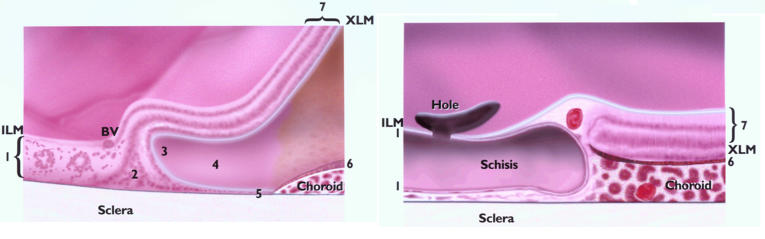
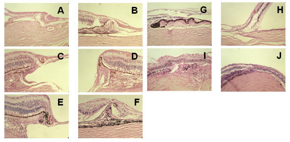
Results: In children, colobomas were associated with lethal malformations. The extracolobomatous inner retinal layers extended centrally, forming the intercalary membrane. Duplication of the outer retinal layers and a horizontal shift of Müllerian glia created a triangle and a locus minoris resistentiae adjacent to the laterally displaced pigment epithelium. Part of the locus was an incomplete layer of photoreceptors excluding Müllerian glia. In adults, atrophy of the intercalary membrane, manifested as central schisis, thinning of the neuroepithelium, and hole formation, was related to a paucity of blood vessels within and underneath the intercalary membrane and the size of colobomas. The margins featured blood vessels, pigment epithelial hypertrophy, and choroidal and scleral thickening in a compact, intertwined arrangement.

Conclusions: Glial atrophy, schisis, and hole formation in the intercalary membrane and separation of the locus minoris resistentiae from the pigment epithelium can disrupt barriers to fluid flow and set the stage for rhegmatogenous retinal detachment. That process is exacerbated by scleral ectasia, increasing vitreous traction at the margin, and retinovascular ischemia within the intercalary membrane. A vascularized, compact margin resembling a laser barrier, found predominantly in adults, may protect against retinal detachment.


Figures

FIGURE 1
FIGURE 1
Schematic comparison of the margin of pediatric (left) and adult (right) colobomas. 1 = intercalary membrane, 2 = glial triangle, 3 = point of reversal, 4 = duplication, 5 = locus minoris resistentiae, 6 = pigmented epithelium, 7 = extracolobomatous retina. Young colobomas are characterized by features 1 through 7, adult by 1, 6, and 7. The absence of 2 through 5 possibly results in a more resilient barrier.
FIGURE 2
FIGURE 2
Histologic findings at the center of colobomas in pediatric patients. In general, the retinal pigment epithelium and choroid are lacking and the retina (intercalary membrane) is directly apposed to the sclera. Special features are rosette formation (A, B), schisis (D), and variable neuronal atrophy (E, F). A, Patient 6, hematoxylin-eosin, ×10. B, Patient 20, hematoxylin-eosin, ×10. C, Patient 11, hematoxylin-eosin, ×10. D, Patient 1, hematoxylin-eosin, ×4. E, Patient 20, hematoxylin-eosin, ×4. F, Patient 20, hematoxylin-eosin, ×40.
FIGURE 3
FIGURE 3
Histologic examination of the margins of colobomas in pediatric patients. In general, the inner retinal layer extends centrally, and the outer layer reverts to meet the laterally displaced retinal pigment epithelium (RPE). The perverse orientation of the photoreceptors is shown (A, B, C, E), as are the radial glia oriented parallel to scleral fibers forming a triangle between the inner and outer retina (D, E, F, I). There is exceptional vascular support of the margin (D), vitreous traction (G), and schisis of the transition to intercalary membrane (H). There is choroidal thickening and RPE hyperplasia (I, J), as well as subchoroidal invasion by the retina (I), which resembles a pocket. Rosettes are part of the margin (A, C). A, Patient 6, hematoxylin-eosin, ×10. B, Patient 21, hematoxylin-eosin, ×10,. C, Patient 18, hematoxylin-eosin, ×10. D, Patient 20, hematoxylin-eosin, ×10. E, Patient 18, hematoxylin-eosin, ×4. F, Patient 23, hematoxylin-eosin, ×10. G, Patient 2, hematoxylin-eosin, ×4. H, Patient 1, hematoxylin-eosin, ×4. I, patient 11, hematoxylin-eosin, ×10. J, Patient 20, hematoxylin-eosin, ×10.
FIGURE 4
FIGURE 4
Histologic examination of the center of colobomas in adult patients. In general, the variably atrophic intercalary membrane is directly apposed to the sclera. Neuronal atrophy (A through J), schisis (D, I), and arteriosclerosis of intercalary vessels (G through J) are consistent with aging. The thinned sclera contains nerves (E, F). A, Patient 7, hematoxylin-eosin, ×10. B, Patient 8, hematoxylin-eosin, ×10. C, Patient 8, hematoxylin-eosin, ×40. D, Patient 14, hematoxylin-eosin, ×10. E, Patient 26, hematoxylin-eosin, ×10. F, Patient 14, hematoxylin-eosin, ×40. G, Patient 8, hematoxylin-eosin, ×10. H, Patient 8, hematoxylin-eosin, ×40. I, Patient 14, hematoxylin-eosin, ×10. J, Patient 22, periodic acid–Schiff stain, ×10.
FIGURE 5
FIGURE 5
Histologic examination of the margin in adult patients. In general, there is an abrupt termination of the retinal pigment epithelium and choroid, which are often hyperplastic and thickened. Both are centrally displaced, eliminating the point of reversal and locus minoris resistentiae and thus adding to structural stability. Moreover, abundant vascular supply of the margin is seen (A, C, D, E). Choroidal thickening and pockets anchor the intercalary membrane and form a seal (B through F), and intercalary schisis and hole formation reduce traction on the margin (C, E). Drusen-like deposits are consistent with aging (G). In smaller colobomas, well-vascularized intercalary membranes show preservation of inner layers (I, J). A, Patient 7, hematoxylin-eosin, ×10. B, Patient 8, hematoxylin-eosin, ×4. C, Patient 8, hematoxylin-eosin, ×10. D, Patient 8, hematoxylin-eosin, ×10. E, Patient 8, hematoxylin-eosin, ×10. F, Patient 14, hematoxylin-eosin, ×4. G, Patient 22, hematoxylin-eosin, ×10. H, Patient 15, hematoxylin-eosin, ×10. I, Patient 9, hematoxylin-eosin, ×4. J, Patient 26, hematoxylin-eosin, ×10.

References

    1. Von Ammon. Anatomische Untersuchung von Coloboma bulbi. Z Ophthalmol 1830: I.
    1. Jesberg D, Schepens C. Retinal detachment associated with coloboma of the choroid. Arch Ophthalmol. 1961;65:163–173. - PubMed
    1. Schepens CL. Retinal Detachment and Allied Diseases Vol 2. Philadelphia: WB Saunders; 1983:615–617.
    1. Hannover A. Cited by Hess C. Zur Pathogenese des Microphthalmus. Archiv Ophthalmol. 1888;34:162.
    1. Hannover A. Funiculus sclerae. Séance de L’academie des sciences danoise. 8. XII. 1876.

Publication types

MeSH terms