. 2005:103:537-67.

Perifoveal vitreous detachment and its macular complications

Affiliations

Perifoveal vitreous detachment and its macular complications

Mark W Johnson. Trans Am Ophthalmol Soc. 2005.

Abstract

Purpose: To evaluate the vitreoretinal relationship in eyes with idiopathic macular disorders putatively caused by vitreomacular traction associated with early stages of age-related posterior vitreous detachment (PVD).

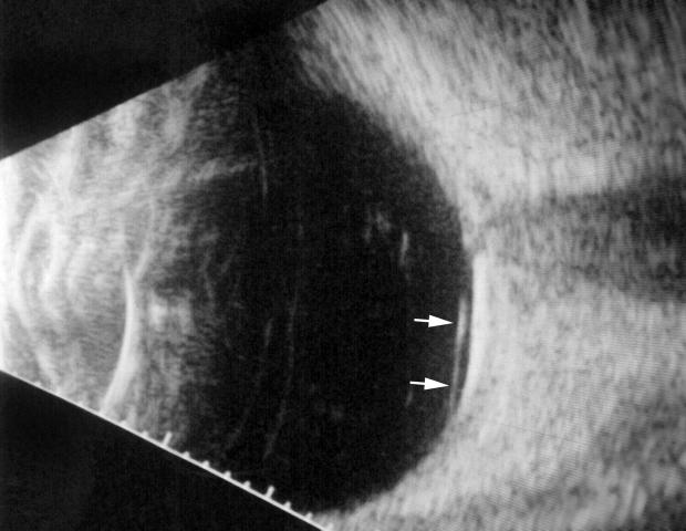
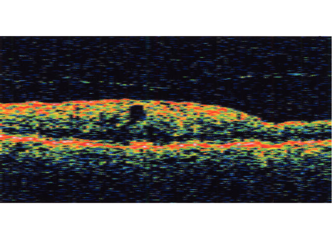
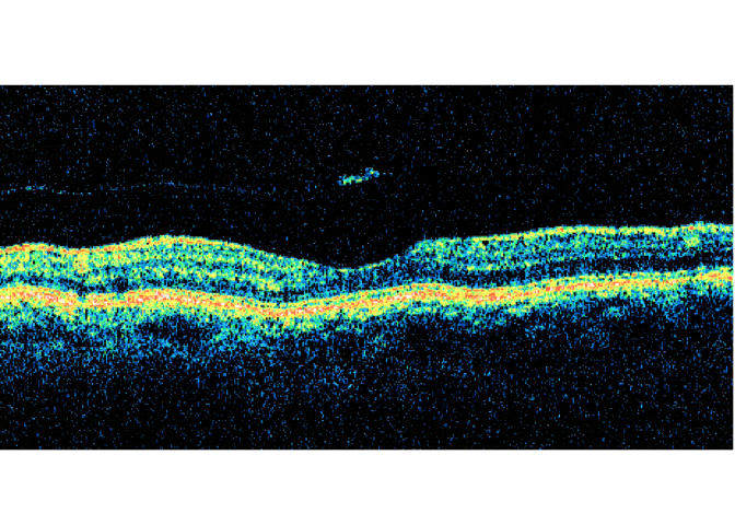
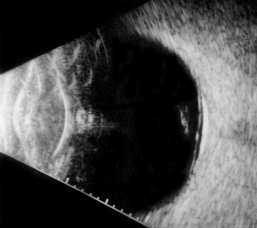
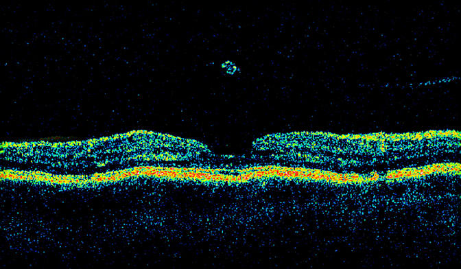
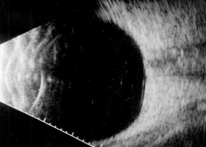
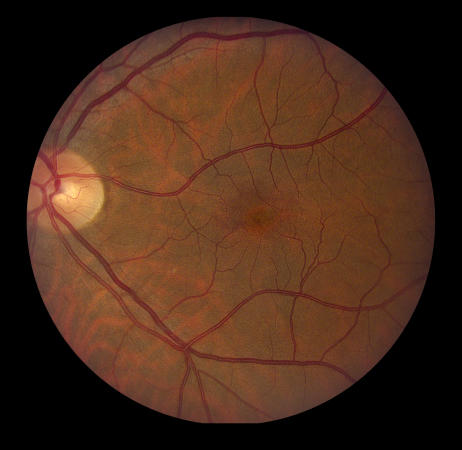
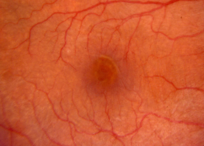
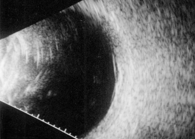
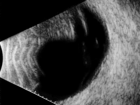
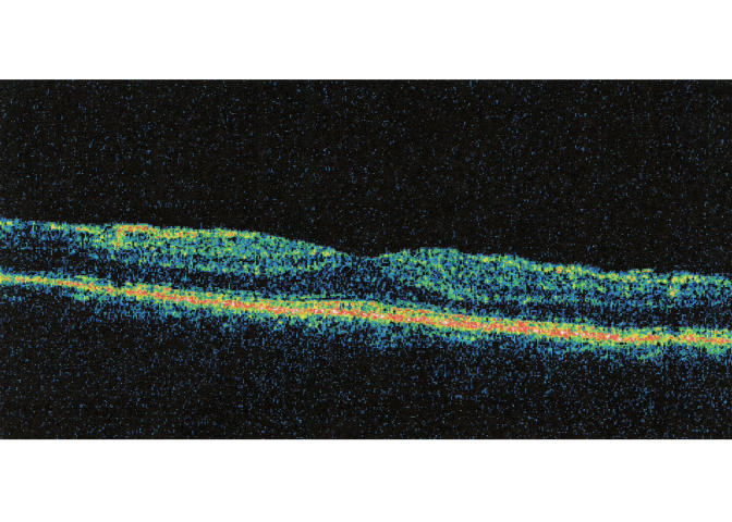
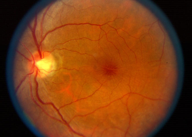
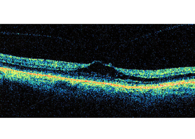
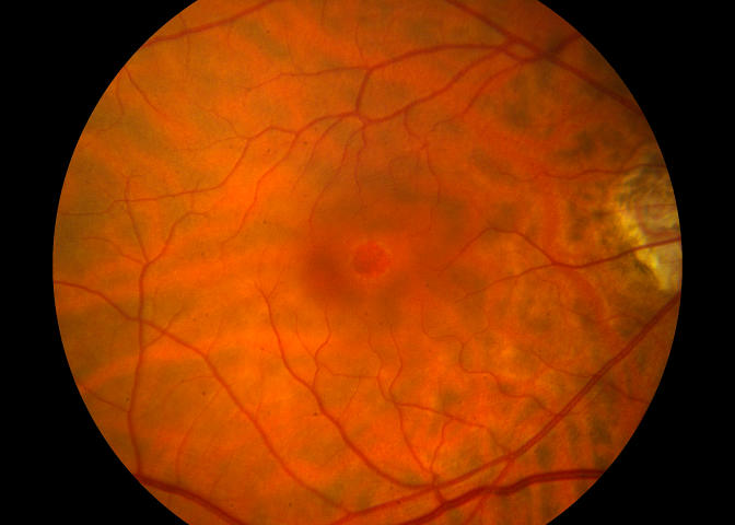
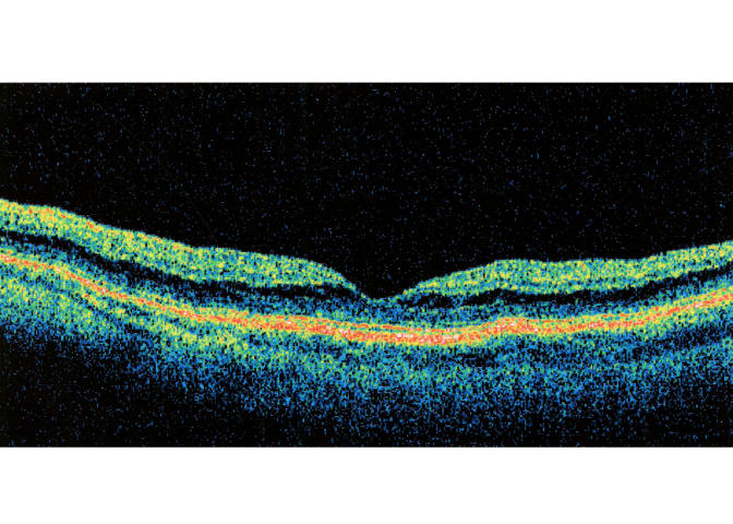
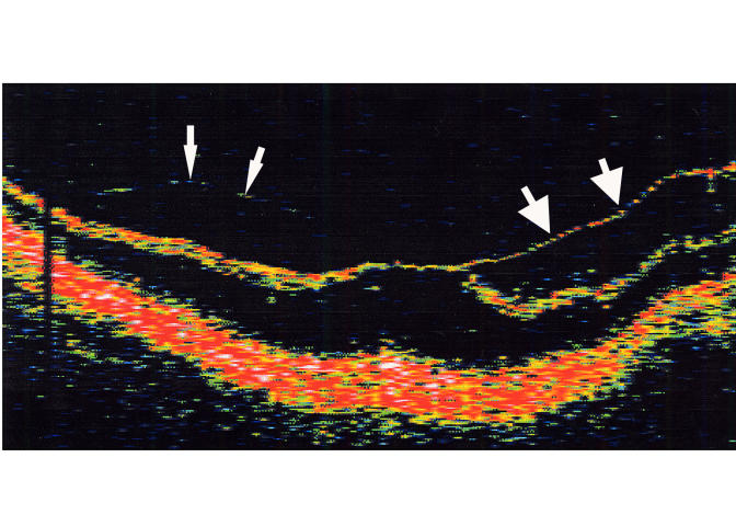
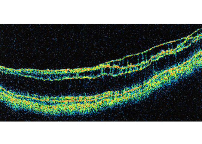
Methods: Retrospective observational case series of 43 eyes of 40 patients diagnosed with one of several idiopathic vitreomacular conditions. Included patients had no biomicroscopic evidence for complete PVD on presentation and underwent a specific clinical assessment of the vitreoretinal relationship. Affected eyes were evaluated with slit-lamp biomicroscopy, B-scan ultrasonography, optical coherence tomography, and/or intraoperative examination of the posterior hyaloid.

Results: By one or more examination techniques, 41 (95.3%) of the 43 study eyes had evidence of vitreous detachment from the perifoveal macular region and the remaining two eyes had complete PVD. When measurable, the size of the vitreomacular adhesion varied by diagnosis. Of 31 eyes with perifoveal vitreous detachment seen in follow-up, only three (9.7%) showed progression to complete PVD over an average preoperative or total follow-up period of 30.0 months (range, 2 to 237 months). Surgical or spontaneous separation of the residual vitreomacular adhesion in 16 eyes was followed in 15 (93.8%) by partial or complete resolution of the symptoms and signs of macular traction.

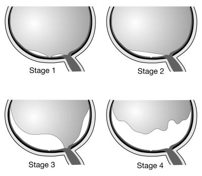
Conclusions: Age-related PVD appears to be an insidious, chronic event that begins in the perifoveal macula and evolves over a prolonged period of time prior to vitreopapillary separation. Though usually asymptomatic, its early (perifoveal) stages may be complicated by one of several macular pathologies, determined in part by the size of the residual vitreomacular adhesion.


Figures

FIGURE 1A
FIGURE 1A
Horizontal axial B-scan ultrasonogram of a localized, shallow detachment of the posterior hyaloid membrane (arrows) from the macular area.
FIGURE 1B
FIGURE 1B
Transverse ultrasound image of total posterior vitreous detachment.
FIGURE 2
FIGURE 2
Schematic illustration of the stages of posterior vitreous detachment. Stage 1, perifoveal vitreous detachment with residual vitreofoveal adhesion. Stage 2, perifoveal vitreous detachment with no vitreofoveal adhesion. Stage 3, near-complete posterior vitreous detachment (PVD) with only vitreopapillary adhesion remaining. Stage 4, complete PVD.
FIGURE 3
FIGURE 3
Case 2. Optical coherence tomography scan of stage 1 perifoveal vitreous detachment associated with a small grade 1 epiretinal membrane (not seen in this meridian).
FIGURE 4A
FIGURE 4A
Case 8. Fundus photograph of right eye showing grade 3 epiretinal membrane (ERM) associated with mild vascular distortion and mild foveal prolapse through central defect in ERM.
FIGURE 4B
FIGURE 4B
Case 8. Horizontal optical coherence tomography image showing stage 1 perifoveal vitreous detachment with vitreous attachment to prolapsing foveola.
FIGURE 5A
FIGURE 5A
Case 10. Fundus photograph of right eye showing grade 3 epiretinal membrane with pseudohole and no biomicroscopic evidence for posterior vitreous detachment.
FIGURE 5B
FIGURE 5B
Case 10. Vertical axial ultrasound image demonstrating shallow stage 2 vitreous detachment over the macular region.
FIGURE 5C
FIGURE 5C
Case 10. Optical coherence tomography confirms shallow separation of the posterior hyaloid overlying epiretinal membrane and cystoid pseudohole.
FIGURE 6
FIGURE 6
Case 12. Optical coherence tomography scan of pseudo-operculum suspended on detached posterior hyaloid over normal fovea.
FIGURE 7
FIGURE 7
Case 15. Vertical axial B-scan ultrasonogram shows shallow stage 2 vitreous detachment with pseudo-operculum. The underlying fovea was normal on biomicroscopy.
FIGURE 8A
FIGURE 8A
Case 17. Fundus photograph of right eye demonstrating lamellar macular hole with overlying operculum.
FIGURE 8B
FIGURE 8B
Case 17. Composite optical coherence tomography image showing small operculum suspended on minimally reflective posterior hyaloid over inner lamellar foveal defect.
FIGURE 9A
FIGURE 9A
Case 18. Fundus photograph of right eye showing macular microhole in patient with acute onset of a tiny central scotoma.
FIGURE 9B
FIGURE 9B
Case 18. Horizontal axial ultrasound of right eye demonstrating shallow detachment of posterior hyaloid over macular region.
FIGURE 9C
FIGURE 9C
Case 18. Fundus photograph of right eye 3 months later shows spontaneously healed microhole.
FIGURE 9D
FIGURE 9D
Case 18. Fundus photograph of left eye 48 months after the development of microhole shows tiny persistent red spot centrally with no apparent foveal defect. Patient still has tiny scotoma subjectively.
FIGURE 9E
FIGURE 9E
Case 18. Optical coherence tomography of left eye shows healed microhole and persistence of stage 2 vitreous detachment 48 months after onset.
FIGURE 10A
FIGURE 10A
Case 22. Macular photograph of right eye showing cystoid macular thickening with shallow ridge of inner retinal traction along superonasal edge of foveola.
FIGURE 10B
FIGURE 10B
Case 22. Vertical macular ultrasound image showing shallow perifoveal vitreous detachment with residual foveolar adhesion.
FIGURE 11A
FIGURE 11A
Case 26. Vertical optical coherence tomography scan of left eye shows perifoveal hyaloidal detachment with focal foveolar attachment causing tractional cystoid foveal thickening. On biomicroscopy, the detached posterior hyaloid was invisible initially and became visible over 2-year follow-up period.
FIGURE 11B
FIGURE 11B
Case 26. Longitudinal ultrasound view of same eye, showing limited extent of perifoveal vitreous detachment.
FIGURE 12A
FIGURE 12A
Case 27. Vertical optical coherence tomography image of left eye shows pagoda-shaped, cystoid foveal thickening and mild epiretinal membrane associated with perifoveal vitreous detachment. Note the increased thickening of posterior hyaloid membrane near its attachment to the foveola.
FIGURE 12B
FIGURE 12B
Case 27. Vertical ultrasound image shows perifoveal vitreous separation, slightly more extensive superiorly.
FIGURE 12C
FIGURE 12C
Case 27. Optical coherence tomography scan 6 months postoperatively shows resolution of tractional cystoid macular edema.
FIGURE 13A
FIGURE 13A
Case 29. Fundus photograph of left eye shows cystoid foveal thickening and radial inner retinal striae. The posterior hyaloid was not visible on biomicroscopy.
FIGURE 13B
FIGURE 13B
Case 29. Corresponding midphase fluorescein angiogram reveals no leakage from retinal capillaries.
FIGURE 13C
FIGURE 13C
Case 29. Optical coherence tomography scan demonstrates cystoid foveal thickening associated with perifoveal vitreous detachment.
FIGURE 13D
FIGURE 13D
Case 29. Optical coherence tomography scan 20 months later shows only slight progression of vitreous separation and associated tractional deformation of the fovea.
FIGURE 14
FIGURE 14
Schematic illustration of vitreomacular interface encountered in cases of tractional cystoid macular edema accompanied by mild epiretinal membrane (ERM). A, The vitreous is firmly anchored to the fovea by extension of ERM onto the posterior hyaloid. This prevents intraoperative separation of vitreous from the fovea from the surgical plane shown in frame B. C, Vitreofoveal separation occurs readily upon peeling the associated ERM.
FIGURE 15A
FIGURE 15A
Ultrasound image of vitreomacular traction syndrome case showing peripheral vitreous detachment with broad area of vitreomacular adhesion. Vertical transverse scan of right eye, case 32.
FIGURE 15B
FIGURE 15B
Ultrasound image of vitreomacular traction syndrome case showing peripheral vitreous detachment with broad area of vitreomacular adhesion. Longitudinal scan of right eye, case 34.
FIGURE 16
FIGURE 16
Case 33. Fundus photograph of left eye with classic vitreomacular traction syndrome. There is vitreous attachment to a broad area involving the macula and optic disc margin, associated with shallow traction macular detachment and epiretinal membrane.
FIGURE 17A
FIGURE 17A
Case 39. Fundus photograph reveals deep yellow foveal ring suggesting outer foveolar defect (stage 1B hole) associated with schisis-like retinal thickening throughout the macular region.
FIGURE 17B
FIGURE 17B
Case 39. Corresponding optical coherence tomography image shows perifoveal vitreous detachment, break in the outer foveola, and outer retinoschisis throughout the macula.
FIGURE 17C
FIGURE 17C
Case 39. Three-month postoperative fundus photograph demonstrates resolution of both macular hole and retinoschisis.
FIGURE 17D
FIGURE 17D
Case 39. Postoperative optical coherence tomography demonstrates resolution of both macular hole and retinoschisis.
FIGURE 18A
FIGURE 18A
Case 40. Fundus photograph shows posterior staphyloma in high myopia. Biomicroscopy revealed retinal thickening throughout the area of the staphyloma.
FIGURE 18B
FIGURE 18B
Case 40. Preoperative horizontal optical coherence tomography shows extensive outer retinoschisis, with an area of inner retinoschisis temporally (large arrows). Detachment of the posterior hyaloid is seen nasally (small arrows).
FIGURE 18C
FIGURE 18C
Case 40. One year postoperatively, optical coherence tomography shows persistence of both inner and outer retinoschisis.

References

    1. Sebag J. The Vitreous: Structure, Function, and Pathobiology. New York: Springer-Verlag; 1989:80–95.
    1. Jaffe NS. Complications of acute posterior vitreous detachment. Arch Ophthalmol. 1968;79:568–571. - PubMed
    1. Foos RY, Wheeler NC. Vitreoretinal juncture: synchysis senilis and posterior vitreous detachment. Ophthalmology. 1982;89:1502–1512. - PubMed
    1. Eisner G. Posterior vitreous detachment. Klin Monatsbl Augenheilkd. 1989;194:389–392. - PubMed
    1. Balazs EA, Denlinger JL. Aging changes in the vitreous. In: Sekuler R, Kline D, Dismukes K, eds. Aging and Human Visual Function. New York: Liss; 1982:45–57.

MeSH terms