. 2006 Nov;44(5):1231-9.
doi: 10.1002/hep.21366.

Cellular and humoral autoimmunity directed at bile duct epithelia in murine biliary atresia

Affiliations

Cellular and humoral autoimmunity directed at bile duct epithelia in murine biliary atresia

Cara L Mack et al. Hepatology. 2006 Nov.

Abstract

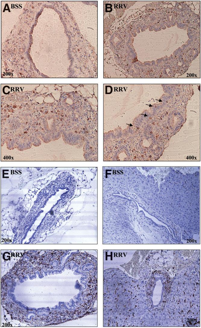
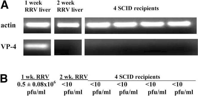
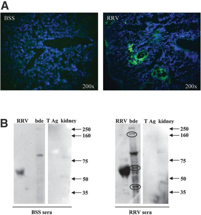
Biliary atresia is an inflammatory fibrosclerosing lesion of the bile ducts that leads to biliary cirrhosis and is the most frequent indication for liver transplantation in children. The pathogenesis of biliary atresia is not known; one theory is that of a virus-induced, subsequent autoimmune-mediated injury of bile ducts. The aim of this study was to determine whether autoreactive T cells and autoantibodies specific to bile duct epithelia are present in the rotavirus (RRV)- induced murine model of biliary atresia and whether the T cells are sufficient to result in bile duct inflammation. In vitro analyses showed significant increases in IFN-gamma-producing T cells from RRV-diseased mice in response to bile duct epithelial autoantigen. Adoptive transfer of the T cells from RRV-diseased mice into naïve syngeneic SCID recipients resulted in bile duct-specific inflammation. This induction of bile duct pathology occurred in the absence of detectable virus, indicating a definite response to bile duct autoantigens. Furthermore, periductal immunoglobulin deposits and serum antibodies reactive to bile duct epithelial protein were detected in RRV-diseased mice. In conclusion, both cellular and humoral components of autoimmunity exist in murine biliary atresia, and the progressive bile duct injury is due in part to a bile duct epithelia-specific T cell-mediated immune response. The role of cellular and humoral autoimmunity in human biliary atresia and possible interventional strategies therefore should be the focus of future research.


Figures

Fig. 1
Fig. 1
Bile duct epithelial antigen–specific autoreactive T cells present in 2-week-old Rhesus rotavirus (RRV)-diseased mice. Purified T cells (rested with IL-15) from livers (A) or spleens (B) of 2-week-old mice previously inoculated with balanced salt solution (BSS) or RRV (responders, 2 × 10⁵ cells/well) were cultured for 48 hours with mitomycin-treated naïve splenocytes (stimulators, 5 × 10⁵ cells/well) in media alone or with antigen. Determination of activation was performed by measuring T cell production of interferon gamma (IFN-γ) using a standard ELISPOT technique. Both RRV-liver and RRV-spleen T cells produced significant amounts of IFN-γ in the presence of bile duct epithelia (bde) homogenate in a dose–response fashion, in comparison with no antigen control (*P < .05). Increased amounts of IFN-γ also were detected in cultures that contained inactivated RRV antigen, suggesting the presence of memory T cells to virus or molecular mimicry. Low IFN-γ was seen within the following controls: media alone (no antigen), kidney epithelia homogenate, SV40 antigen, antigen-presenting cells (APCs) alone, and T cells alone. Furthermore, BSS-liver and BSS-spleen T cells produced low/undetectable IFN-γ in response to multiple antigens. All responders (BSS and RRV groups) produced excess amounts of IFN-γ (TNTC) in the presence of phytohemagglutinin (PHA) (positive control; not shown).
Fig. 2
Fig. 2
CD4+ T cell blockade abrogates interferon gamma (IFN-γ) production from T cells of Rhesus rotavirus (RRV)-diseased mice stimulated with bile duct epithelial antigen. Purified CD3+ T cells (rested with IL-15) from spleens of 2-week-old mice previously inoculated with RRV (responders, 4 × 10⁵ cells/well) were cultured for 48 hours with mitomycin-treated naïve splenocytes (stimulators, 5 × 10⁵ cells/well) with bile duct epithelial antigen [100 μg/mL bile duct epithelial homogenate (bde)] alone or in combination with anti-CD4, anti-CD8, or IgG2b (isotype control). Determination of activation was performed by measuring T cell production of IFN-γ using the ELISPOT technique. Marked suppression of T cells producing IFN-γ was observed in the presence of CD4 blocking antibody. This suppression was not found in cultures with CD8 blocking antibody or isotype control.
Fig. 3
Fig. 3
SCID recipients of adoptively transferred liver T cells from Rhesus rotavirus (RRV)-diseased mice have prominent periductal infiltrates. Adult SCID C.B-17 mice received 4 to 5 × 10⁵ liver T cells or 8 × 10⁵ spleen T cells from 2-week-old balanced salt solution (BSS)-inoculated or RRV-diseased mice. Shown here are representative hematoxylineosin–stained sections from the extrahepatic ducts and porta hepatis regions of SCID mice, 2 weeks after adoptive transfer. Black arrows point to bile ducts. The SCID mice that received BSS liver T cells did not have periductal infiltrates of the extrahepatic bile ducts (A) or large intrahepatic ducts (B). In contrast, SCID recipients of RRV liver T cells (C-D) or spleen T cells (not shown) had prominent immune cell infiltrates surrounding both the extrahepatic ducts and large intrahepatic ducts. No evidence of inflammation was seen within or surrounding the ductal epithelia of pancreatic ducts or renal tubular epithelia in SCID recipients of donor T cells from RRV-diseased mice (Fig. 2E-F).
Fig. 4
Fig. 4
Periductal infiltrates within SCID recipients of Rhesus rotavirus (RRV) liver T cells contained the adoptively transferred donor CD3+ T cells and recipient macrophages. Representative sections from SCID recipients of balanced salt solution (BSS) control or RRV liver T cells were stained for antibody to CD3 or F4/80 (shown in brown) to identify adoptively transferred T cells and recipient macrophages, respectively. Few CD3+ T cells were seen surrounding the extrahepatic bile ducts in SCID recipients of BSS liver T cells (A). In contrast, the extrahepatic bile duct epithelia from SCID recipients of RRV liver T cells were surrounded by an abundance of CD3+ T cells (B-C; 400× magnified view). T cells also were found invading in between the bile duct epithelia (D, 400× ). SCID recipients of BSS liver T cells did not have F4/80+ cells (macrophages) surrounding either the extrahepatic bile ducts (E) or large intrahepatic ducts (F) In contrast, a marked influx of recipient macrophages was observed surrounding the extrahepatic ducts (G) and large intrahepatic ducts (H) in SCID recipients of RRV liver T cells. Note the F4/80+ staining of sinusoidal macrophages in the parenchyma of both groups.
Fig. 5
Fig. 5
Absence of mRNA expression of Rhesus rotavirus (RRV) structural protein VP-4 and actively replicating virus in donor RRV-liver T cell preparations or recipient SCID mice livers. (A) Reverse transcription polymerase chain reaction (RT-PCR): RNA from 1-week-old liver homogenates of RRV-diseased mice (positive control), pooled donor liver immune cells (Percoll isolation) from 2-week-old RRV-diseased mice, and liver homogenates from four SCID recipients of RRV-diseased liver T cells were extracted and used in RT-PCR reactions for actin (control) and VP-4 (structural protein of RRV). PCR evidence of RRV RNA was found in the 1-week-old RRV-diseased liver homogenates but absent in the donor T cell preparations from 2-week-old RRV-diseased mice as well as SCID recipients, 2 weeks after adoptive transfer of RRV liver T cells. (B) Infectious plaque assay: One-week-old RRV liver homogenates (positive control) had 0.5 × 10⁶ ± 0.08 × 10⁶ pfu/mL infectious virus whereas undetectable levels (<10 pfu/mL) of infectious virus were observed in the 2-week-old RRV-liver donor T cells and recipient SCID liver homogenates.
Fig. 6
Fig. 6
Peri-ductal IgG immune deposits and serum antibodies reactive to bile duct epithelial proteins are evident in 2-week-old Rhesus rotavirus (RRV)-diseased mice. Liver tissue and serum from 2-week-old mice previously inoculated with balanced salt solution (BSS) or RRV were obtained for immunohistochemistry and Western blot analysis, respectively. (A) Indirect immunofluorescence: Liver sections were analyzed for the presence of IgG immune deposits on bile duct epithelia by standard immunofluorescent techniques. Tissue was incubated with goat anti-mouse IgG-fluorescein isothiocyanate (FITC) (green) and counterstained stained with Hoechst dye (blue) to detect cellular nuclei. RRV-diseased mouse livers had detectable IgG immune deposits surrounding bile duct epithelia within portal tracts. No detectable IgG deposits were seen in livers from BSS control mice. (B) Western blot analysis: Irradiated RRV and bile duct epithelial cell line homogenate (and controls SV40 large T antigen and kidney epithelia homogenate) were separated by gel electrophoresis, blotted onto nitrocellulose paper, and incubated with either BSS or RRV sera. Antibodies reactive to protein were visualized by chemiluminescence (dark bands on radiograph). Both the BSS and RRV sera contained nonspecific antibodies reactive to (1) a viral protein at approximately 60 kd molecular weight; (2) bile duct epithelial proteins of approximately 80 kd and 250 kd; and (3) kidney epithelial proteins of 50 kd and 80 kd. Neither sera from BSS or RRV reacted with large T antigen. Importantly, the RRV sera, and not the BSS sera, contained antibodies reactive to multiple bile duct epithelial proteins, including strong reactivity to proteins of approximately 40, 60, and 160 kd (circled bands). These bands represent novel bile duct epithelial autoantigens bound by serum antibodies from 2-week-old RRV-diseased mice.

References

    1. Mack CL, Tucker RM, Sokol RJ, Karrer FM, Kotzin BL, Whitington PF, et al. Biliary atresia is associated with CD4+ Th1 cell-mediated portal tract inflammation. Pediatr Res. 2004;56:79–87. - PMC - PubMed
    1. Davenport M, Gonde C, Redkar R, Koukoulis G, Tredger M, Mieli-Vergani G, et al. Immunohistochemistry of the liver and biliary tree in extrahepatic biliary atresia. J Pediatr Surg. 2001;36:1017–1025. - PubMed
    1. Tracy TF, Dillon P, Fox ES, Minnick K, Vogler C. The inflammatory response in pediatric biliary disease: macrophage phenotype and distribution. J Pediatr Surg. 1996;31:121–125. - PubMed
    1. Kobayashi H, Puri P, O'Briain DS, Surana R, Miyano T. Hepatic overexpression of MHC class II antigens and macrophage-associated antigens (CD68) in patients with biliary atresia of poor prognosis. J Pediatr Surg. 1997;32:590–593. - PubMed
    1. Karrer FM, Price MR, Bensard DD, Sokol RJ, Narkewicz MR, Smith DJ, et al. Long-term results with the Kasai operation for biliary atresia. Arch Surg. 1996;131:493–496. - PubMed

Publication types

MeSH terms