. 2006 Oct 31;45(43):12917-28.
doi: 10.1021/bi060717k.

The crystal structure of the BAR domain from human Bin1/amphiphysin II and its implications for molecular recognition

Affiliations

The crystal structure of the BAR domain from human Bin1/amphiphysin II and its implications for molecular recognition

Eva Casal et al. Biochemistry. .

Abstract

BAR domains are found in proteins that bind and remodel membranes and participate in cytoskeletal and nuclear processes. Here, we report the crystal structure of the BAR domain from the human Bin1 protein at 2.0 A resolution. Both the quaternary and tertiary architectures of the homodimeric Bin1BAR domain are built upon "knobs-into-holes" packing of side chains, like those found in conventional left-handed coiled-coils, and this packing governs the curvature of a putative membrane-engaging concave face. Our calculations indicate that the Bin1BAR domain contains two potential sites for protein-protein interactions on the convex face of the dimer. Comparative analysis of structural features reveals that at least three architectural subtypes of the BAR domain are encoded in the human genome, represented by the Arfaptin, Bin1/Amphiphysin, and IRSp53 BAR domains. We discuss how these principal groups may differ in their potential to form regulatory heterotypic interactions.


Figures

Figure 1
Figure 1
Overall structure of the BAR domain of Bin1 A, Protomer of Bin1BAR. The numbering refers to the helices, starting from the N terminus. B, The Bin1 homo-dimer, generated from crystallographic symmetry. The protein forms a banana-shaped homo-dimer. C, View of the concave face of the dimer. D, View of the convex face of the dimer.
Figure 2
Figure 2
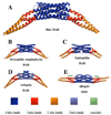
Ribbon representation of the different BAR domains of known structure A, The helical arrangement in Bin1BAR consists of a central 6-helix bundle (blue), flanked by a 3-helix bundle (orange-red) and, finally, a 2-helix coiled-coil at the periphery (yellow). B, Drosophila Amphiphysin BAR and C, Endophilin BAR share the same helical arrangement. D, Arfaptin BAR contains a region that corresponds to a 5-helix bundle (violet). E, IRSp53 IMD contains an extra helix (green) that associates with the 3-helix bundle (orange-red).
Figure 3
Figure 3
Electrostatic potential surface analysis of Bin1BAR using the program GRASP (32). Bin1BAR shows the canonical bipartite charge distribution present in other BAR domains: positive charge in the concave face (left), negative charge in the convex face (right). The dashed circles indicate the positively charged residues that were mutated in Amphiphysin II and rendered the BAR domain unable to bind to liposomes (2).
Figure 4
Figure 4
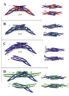
Comparison of the curvature of Bin1BAR with other BAR domains of known structure. Curvatures were compared by superposing the structure of Bin1BAR (navy blue) with all the other structures. The numbers i, ii and iii correspond to the side, concave and convex view of the dimer respectively. Number iv corresponds to the superpositions of the protomers alone, where appropriate. A, Bin1BAR dimer (blue) superposed with Drosophila Amphiphysin BAR (red). B, Bin1BAR dimer (blue) superposed with Endophilin BAR (grey). C, Bin1BAR dimer (blue) superposed with Arfaptin 2 BAR (pink). D, Bin1BAR (blue) superposed with the IMD domain of IRSp53 (green).
Figure 5
Figure 5
Optimal docking area (ODA) analysis of Bin1BAR and Arfaptin2 BAR. The difference between the proteins indicates that Bin1BAR is unlikely to engage in interactions with G-proteins. Numbers i/, ii/ and iii/ correspond to the concave, side and convex view of the dimer respectively. A, ODA analysis for a Bin1BAR protomer. As can be seen from the dark blue patches, the dimerisation interface in the protomer is clearly predicted with this analysis. B, ODA analysis for the Bin1BAR dimer. The concave face of the Bin1BAR dimer has no predicted propensity for protein interaction. C, ODA analysis for the Arfaptin2 BAR dimer. D, Structure of Arfaptin2 BAR (white) in complex with Rac (blue). A comparison of C and D clearly indicates that the binding site for Rac in Arfaptin is correctly predicted by the ODA calculations.
Figure 6
Figure 6
Optimal docking area (ODA) analysis of BAR domain structures and of the IMD domain structure of IRSp53. Numbers i/, ii/ and iii/ correspond to the concave, side and convex view of the dimer respectively. A, Bin1BAR, B, Drosophila Amphiphysin BAR, C, Endophilin BAR, D, Arfaptin BAR and E,IMD domain of IRSp53. BAR domains from A, to D, possess a predicted site with potential for protein-protein interactions on the convex face of the dimer (shown as arrows in iii). For some BAR domains, this potential is strong, such as in endophilin BAR and Arfaptin BAR (C and D) while in the amphiphysins (A and B) it is only weak. This site does not seem to be present in the IMD domain of IRSp53 (E). However, a site analogous to Arfaptin’s Rac interaction site in the concave face is predicted.
Figure 7
Figure 7
A, The structure of the mammalian Amphiphysin II model (dark blue) superimposed to the Bin1 isoform structure (Bin1BAR) (yellow). The modeled insertion region is highlighted with a dashed circle. Numbers i/, ii/ and iii/ correspond to the concave, side and convex view of the dimer respectively. B, Electrostatic analysis of the model. C, ODA analysis of the model.

References

    1. Sakamuro D, Elliott KJ, Wechsler-Reya R, Prendergast GC. BIN1 is a novel MYC-interacting protein with features of a tumour suppressor. Nat Genet. 1996;14:69–77. - PubMed
    1. Peter BJ, Kent HM, Mills IG, Vallis Y, Butler PJ, Evans PR, McMahon HT. BAR domains as sensors of membrane curvature: the amphiphysin BAR structure. Science. 2004;303:495–499. - PubMed
    1. Tarricone C, Xiao B, Justin N, Walker PA, Rittinger K, Gamblin SJ, Smerdon SJ. The structural basis of Arfaptin-mediated cross-talk between Rac and Arf signalling pathways. Nature. 2001;411:215–219. - PubMed
    1. Millard TH, Bompard G, Heung MY, Dafforn TR, Scott DJ, Machesky LM, Futterer K. Structural basis of filopodia formation induced by the IRSp53/MIM homology domain of human IRSp53. EMBO J. 2005;24:240–250. - PMC - PubMed
    1. Weissenhorn W. Crystal structure of the endophilin-A1 BAR domain. J. Mol. Biol. 2005;351:653–661. - PubMed

Publication types

MeSH terms

Substances