. 2007 Jan;27(1):182-94.
doi: 10.1128/MCB.00846-06. Epub 2006 Oct 23.

Conditional deletion of the mouse Klf4 gene results in corneal epithelial fragility, stromal edema, and loss of conjunctival goblet cells

Affiliations

Conditional deletion of the mouse Klf4 gene results in corneal epithelial fragility, stromal edema, and loss of conjunctival goblet cells

Shivalingappa K Swamynathan et al. Mol Cell Biol. 2007 Jan.

Abstract

The Krüppel-like transcription factor KLF4 is among the most highly expressed transcription factors in the mouse cornea (B. Norman, J. Davis, and J. Piatigorsky, Investig. Ophthalmol. Vis. Sci. 45:429-440, 2004). Here, we deleted the Klf4 gene selectively in the surface ectoderm-derived structures of the eye (cornea, conjunctiva, eyelids, and lens) by mating Klf4-LoxP mice (J. P. Katz, N. Perreault, B. G. Goldstein, C. S. Lee, P. A. Labosky, V. W. Yang, and K. H. Kaestner, Development 129:2619-2628, 2002) with Le-Cre mice (R. Ashery-Padan, T. Marquardt, X. Zhou, and P. Gruss, Genes Dev. 14:2701-2711, 2000). Klf4 conditional null (Klf4CN) embryos developed normally, and the adult mice were viable and fertile. Unlike the wild type, the Klf4CN cornea consisted of three to four epithelial cell layers; swollen, vacuolated basal epithelial and endothelial cells; and edematous stroma. The conjunctiva lacked goblet cells, and the anterior cortical lens was vacuolated in Klf4CN mice. Excessive cell sloughing resulted in fewer epithelial cell layers in spite of increased cell proliferation at the Klf4CN ocular surface. Expression of the keratin-12 and aquaporin-5 genes was downregulated, consistent with the Klf4CN corneal epithelial fragility and stromal edema, respectively. These observations provide new insights into the role of KLF4 in postnatal maturation and maintenance of the ocular surface and suggest that the Klf4CN mouse is a useful model for investigating ocular surface pathologies such as dry eye, Meesmann's dystrophy, and Steven's-Johnson syndrome.


Figures
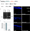
FIG. 1.
FIG. 1.
Conditional deletion of the mouse Klf4 gene. (A) Structure of the Le-Cre and Klf4-LoxP transgenes used. Cre recombinase is under the control of Pax6 P0 promoter and lens/pancreas specific enhancer (top). NLS-Cre, Nuclear localization signal fused to Cre recombinase; IRES-GFP, internal ribosome entry site fused to the gene encoding green fluorescent protein. The Klf4-LoxP transgene contains LoxP sites (triangles) inserted in the first and third introns. In Klf4CN tissues expressing Cre, the second and third exons are excised out, fusing the first exon out of frame with the fourth exon of the Klf4 gene. (B) RT-PCR of Klf4 and RNA polymerase II transcripts in the total RNA from wild-type and Klf4CN corneas. (C) Relative quantities of Klf4 transcripts in the wild-type (WT) and Klf4CN corneas as measured by Q-RT-PCR analysis. (D) Immunohistochemistry with anti-KLF4 antibody in wild-type and Klf4CN corneas. Reactions using wild-type corneas without primary antibody served as controls. Left, DAPI-stained nuclei. Right, fluorescence from secondary antibody against anti-KLF4 antibody.
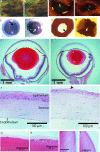
FIG. 2.
FIG. 2.
Morphology and histology of Klf4CN eyes, showing comparisons of wild-type and Klf4CN external appearance (A and B, respectively), eyes (C and D), eyeballs (E and F), dissected cornea and iris viewed from the position of the lens (G and H), midsection of the whole eye (I and J) (magnification, ×25), central cornea (K and L) (magnification, ×400), lens anterior (M and N) (magnification, ×400), and lens equator (O and P) (magnification, ×400). Arrows in panels E and G indicate a well-formed pupil in the wild-type eye; arrows in panels F and H indicate the iris hypertrophy and absence of pupil in the Klf4CN eye. An epithelial bullus in the Klf4CN cornea is indicated (arrowhead in panel L). The whorl-shaped rosette in the wild-type retina to the left of the optic nerve is an artifact of sectioning.
FIG. 3.
FIG. 3.
Ultrastructural analysis of wild-type and Klf4CN corneas. (A and B) Transmission electron microscopy of wild-type (A) and Klf4CN (B) corneas. Klf4CN corneal epithelium contains fewer cell layers, fewer microvilli on the superficial cells (arrows), and swollen, spherical, and vacuolated (arrowheads) basal cells. Delamination at the surface of the Klf4CN corneas is indicated (long arrow in panel B). Magnification: ×5,000. (C to F) Surface scanning electron microscopy at low (C and D) (magnification, ×500) and high (E and F) (magnification, ×10,000) magnifications. Unlike the uniformly stained cells at the wild-type corneal surface (C), the Klf4CN corneal surface contained a mix of electron-dense (arrows in panel D) and light cells.
FIG. 4.
FIG. 4.
Increased cell proliferation in the Klf4CN corneal epithelium. Cryosections of eyeballs from BrdU-injected wild-type and Klf4CN mice probed by immunohistochemistry with anti-BrdU antibody (right panels) are shown. Bright-field images of the corresponding corneal sections are shown on the left.
FIG. 5.
FIG. 5.
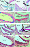
Defective goblet cell development in the Klf4CN conjunctiva. Sections from 10-week-old wild-type (A, C, E, and G) or Klf4CN (B, D, F, and H) mouse heads stained with Alcian blue (A to D) or PAS (E to H) at low (A, B, E, and F) (magnification, ×100) or high (C, D, G, and H) (magnification, ×400) magnification. Dark blue (alcian blue)- or purple (PAS)-stained goblet cell clusters were observed in the wild-type (arrows) but not the Klf4CN conjunctiva. BC, bulbar conjunctiva; PC, palpebral conjunctiva; CF, conjunctival fornix.
FIG. 6.
FIG. 6.
Defects in basement layer deposition in Klf4CN mice. (A) Wild-type (left) or Klf4CN (right) mouse central cornea from midsections stained with PAS. Magnification, ×400. (B) Transmission electron microscopy of thin midsections from the wild-type (left) or Klf4CN (right) mouse central corneas at high magnification (magnification, ×25,000).
FIG. 7.
FIG. 7.
Downregulation of keratin-12 gene expression in the Klf4CN corneal epithelium. (A) Relative quantities of keratin-12 gene transcripts in total RNA from wild-type (WT) and Klf4CN corneas as measured by Q-RT-PCR analysis. Error bars indicate standard deviations. (B) Immunoblot of total proteins from wild-type or Klf4CN cornea, probed with anti-keratin-12 antibody. The blot was stripped and reprobed with antiactin antibody to ensure equal protein loading from wild-type and Klf4CN corneas. (C) Immunohistochemistry with anti-keratin-12 antibody in the wild-type or Klf4CN corneas. Reactions without primary antibody served as controls. Left, DAPI-stained nuclei. Right, fluorescence from secondary antibody against anti-keratin-12 antibody. (D) Relative activity of the Krt12 promoter driving the luciferase reporter gene, measured by cotransfection of increasing amounts of KLF4 expression plasmid pCI-Klf4 in human corneal epithelial cells. (E) Chromatin immunoprecipitation of Krt12 promoter-bound KLF4 by anti-KLF4 antibody, detected by PCR of Krt12 promoter fragment. The negative control is a reaction in which preimmune rabbit serum was used in place of anti-KLF4 antibody.
FIG. 8.
FIG. 8.
Downregulation of aquaporin-5 gene expression in the Klf4CN corneal epithelium. (A) Relative quantities of Aqp5 transcripts in the wild-type (WT) and Klf4CN corneas as measured by Q-RT-PCR analysis. Error bars indicate standard deviations. (B) Immunoblot of total proteins from wild-type or Klf4CN cornea, probed with anti-Aqp5 antibody. The blot was stripped and reprobed with antiactin antibody to ensure equal protein loading from wild-type and Klf4CN corneas. (C). Immunohistochemistry with anti-Aqp5 antibody in the wild-type or Klf4CN corneas. Reactions without primary antibody served as controls. Left, DAPI-stained nuclei. Right, fluorescence from secondary antibody against anti-Aqp5 antibody. D. Relative activity of the Aqp5 promoter driving the luciferase reporter gene, measured by cotransfection of increasing amounts of KLF4 expression plasmid pCI-Klf4 in human corneal epithelial cells. E. Chromatin immunoprecipitation of Aqp5 promoter-bound KLF4 by anti-KLF4 antibody, detected by PCR of Aqp5 promoter fragment. The negative control is a reaction in which preimmune rabbit serum was used in place of anti-KLF4 antibody.

References

    1. Adhikary, G., J. F. Crish, F. Bone, R. Gopalakrishnan, J. Lass, and R. L. Eckert. 2005. An involucrin promoter AP1 transcription factor binding site is required for expression of involucrin in the corneal epithelium in vivo. Investig. Ophthalmol. Vis. Sci. 46:1219-1227. - PubMed
    1. Adhikary, G., J. F. Crish, R. Gopalakrishnan, F. Bone, and R. L. Eckert. 2005. Involucrin expression in the corneal epithelium: an essential role for Sp1 transcription factors. Investig. Ophthalmol. Vis. Sci. 46:3109-3120. - PubMed
    1. Araki-Sasaki, K., Y. Ohashi, T. Sasabe, K. Hayashi, H. Watanabe, Y. Tano, and H. Handa. 1995. An SV40-immortalized human corneal epithelial cell line and its characterization. Investig. Ophthalmol. Vis. Sci. 36:614-621. - PubMed
    1. Argueso, P., M. Balaram, S. Spurr-Michaud, H. T. Keutmann, M. R. Dana, and I. K. Gipson. 2002. Decreased levels of the goblet cell mucin MUC5AC in tears of patients with Sjogren syndrome. Investig. Ophthalmol. Vis. Sci. 43:1004-1011. - PubMed
    1. Ashery-Padan, R., T. Marquardt, X. Zhou, and P. Gruss. 2000. Pax6 activity in the lens primordium is required for lens formation and for correct placement of a single retina in the eye. Genes Dev. 14:2701-2711. - PMC - PubMed

Publication types

MeSH terms