. 2007 Jan;27(1):312-23.
doi: 10.1128/MCB.00985-06. Epub 2006 Oct 23.

The Rac effector p67phox regulates phagocyte NADPH oxidase by stimulating Vav1 guanine nucleotide exchange activity

Affiliations

The Rac effector p67phox regulates phagocyte NADPH oxidase by stimulating Vav1 guanine nucleotide exchange activity

Wenyu Ming et al. Mol Cell Biol. 2007 Jan.

Abstract

The phagocyte NADPH oxidase catalyzes the reduction of molecular oxygen to superoxide and is essential for microbial defense. Electron transport through the oxidase flavocytochrome is activated by the Rac effector p67(phox). Previous studies suggest that Vav1 regulates NADPH oxidase activity elicited by the chemoattractant formyl-Met-Leu-Phe (fMLP). We show that Vav1 associates with p67(phox) and Rac2, but not Rac1, in fMLP-stimulated human neutrophils, correlating with superoxide production. The interaction of p67(phox) with Vav1 is direct and activates nucleotide exchange on Rac, which enhances the interaction between p67(phox) and Vav1. This provides new molecular insights into regulation of the neutrophil NADPH oxidase, suggesting that chemoattractant-stimulated superoxide production can be amplified by a positive feedback loop in which p67(phox) targets Vav1-mediated Rac activation.


Figures
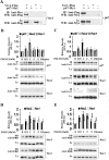
FIG. 1.
FIG. 1.
p67phox interacts with Vav1. (A) COS-7 cells were transiently cotransfected with cDNAs encoding Flag-tagged Vav1 and Myc-tagged p67phox. Immunoprecipitation assays were carried out using Myc (for p67phox) or Flag (for Vav1) MAb and Western blots probed with Flag or Myc MAb as indicated. Each blot is representative of three independent experiments. Human neutrophils (B and D) or myeloid PLB-985 granulocytes (C and E) were treated with 10 μM fMLP for the indicated amounts of time. (B and C) Cell lysates (30 × 106 cells) were immunoprecipitated with a rabbit anti-Vav1 antibody and analyzed by Western blotting with p67phox MAb, rabbit anti-Rac2 antibody, or Rac1 MAb. Enhanced chemiluminescence signal was quantified by densitometry. Bar graphs show mean immunoprecipitation signal normalized to the lysate signal. Assays were carried out in triplicate and the mean ± SD was calculated. (D and E) A GST-PBD pull-down assay was used for quantifying Rac-GTP. Cell lysates were incubated with GST-PBD protein and glutathione-Sepharose beads, and eluted samples were analyzed by immunoblotting using Rabbit anti-Rac2 antibody or Rac1 MAb, as indicated. Assays were carried out in triplicate and the mean ± SD was calculated. Bar graphs showed mean pull-down signal normalized to lysate signal. WB, Western blotting; IP, immunoprecipitation; Ly, whole-cell lysates (1% of the cell lysate used for immunoprecipitation); PD, pull-down assay.
FIG. 2.
FIG. 2.
The interaction of p67phox with Vav1 is direct. Pull-down (PD) samples were analyzed by Western blotting (WB), as indicated. (A) Purified recombinant His-tagged Vav1 (DH-PH-ZF) and GST-p67phox were preincubated in binding buffer prior to addition of glutathione-Sepharose 4B (GSH). GSH pull-down samples were probed with His MAb. (B) Recombinant His-Vav1 (DH-PH-ZF) and GST-p67phox or GST were coincubated prior to the addition of GSH. Pull-down samples were detected with anti-His antibody. (C) His-tagged Vav1 (DH-PH-ZF) and GST-p67phox were preincubated in binding buffer prior to the addition of Ni-NTA agarose beads. Pull-down samples were analyzed with p67phox MAb. These assays were performed in triplicate with similar results.
FIG. 3.
FIG. 3.
p67phox stimulates Vav1 binding to Rac. (A and B) COS-7 cells were transiently cotransfected with cDNAs encoding Vav1 and Myc-tagged Rac1 (A) or Myc-tagged Rac2 (B), with or without Myc-tagged p67phox. Immunoprecipitation samples were prepared with rabbit anti-Vav1 antibody and analyzed by Western blotting, probed with Myc MAb (for p67phox and Rac) or Vav1 MAb. Bar graphs show mean immunoprecipitation Rac signals normalized to lysate Rac signals; a two-tailed Student's t test was used to determine the difference between groups (**, P < 0.0001; n = 5). (C) Immunoprecipitation assays were performed as described for panel A except that cells were lysed in buffer containing 10 mM MgCl2. (D) The immunoblot shown in panel A was stripped and reprobed with Rac1 MAb, showing that endogenous (Endo) and exogenous (Exo) Rac1 were included in Vav1 immunoprecipitation complexes. Results are representative of three independent experiments. IP, immunoprecipitation; Ly, whole-cell lysates prior to immunoprecipitation; WB, Western blotting.
FIG. 4.
FIG. 4.
Rac-GTP stimulates the interaction between p67phox and Vav1. (A) p67phox domains and mutants are shown schematically. TPR, tetracopeptide repeat; AD, active domain; PR, proline-rich region; PB1, Phox and Bem1 domain; N299, fragment comprised of amino acids 1 to 299; C350, truncated N-terminal 350 amino acids of p67phox; R102E and V204A, point mutations. (B to G) COS-7 cells were cotransfected with constructs expressing Vav1 in combination with wild-type or mutant p67phox, wild-type or mutant Rac1, and/or Rho-DGI, as indicated. Immunoprecipitation (IP) assays were performed with rabbit anti-Vav1 polyclonal antibody and Western blots were probed with a Vav1 MAb, p67phox MAb, rabbit anti-Rho-GDI antibody, Rac MAb, or Rac1 MAb, as indicated. Ly, whole-cell lysate.
FIG. 5.
FIG. 5.
Coexpression of Vav1, p67phox and Rac activates endogenous and exogenous Rac. (A and B) GST-PBD assays for Rac activation were carried out as described in the legend of Fig. 1 except that COSphox cells were transfected for expression of wild-type Vav1 and wild-type p67phox or with Rac1 or Rac2 in different combinations; for detection of Rac, immunoblots were probed with either a Rac1 MAb (A) or a Rac1/2 MAb (B). The bar graphs show the relative level of total (endogenous [Endo] plus exogenous [Exo]) activated Rac normalized to total Rac. Assays were performed in triplicate and mean values ± SD are shown. ANOVA followed by a Tukey-Kramer multiple comparison test was used to determine the difference between groups. **, P < 0.001 versus all other groups. (C) GST-PBD assays for Rac1 activation were carried out for wild-type or p67phox(R102E), coexpressed with Vav1 and Rac1 in COS-7 cells. (D and E) In vitro guanine exchange assays were carried out to examine GEF activity of Flag-tagged Vav1 expressed in cotransfected COS-7 cells with or without cotransfection of constructs for expression of p67phox and Rac, as indicated by 3Pl (plasmids for expression of Vav1, p67phox, and Rac), 2Pl (Vav1 and p67phox), or 1Pl (Vav1). Cell lysates were immunoprecipitated by anti-Flag antibody-conjugated beads, and bound proteins were eluted with Flag peptide. The eluted proteins (or elution buffer only) were added to a mixture containing mant-GTP and bacterially expressed GST-Rac at the time indicated by the arrow, and the relative fluorescence of mant-GTP was monitored by a Perkin-Elmer Life Sciences LS 50B spectrophotometer. Labels indicate plasmids used to transfect COS-7 cells prior to immunoprecipitation or addition of buffer alone. The lower group curves in panel E were generated for reaction mixtures lacking GST-Rac1. Assays were performed in triplicate, and each graph represents one experiment. WB, Western blotting; PD, pull-down; Ly, whole-cell lysates.
FIG. 6.
FIG. 6.
Activation of Rac by coexpression of Vav1 and p67phox requires Vav1 GEF activity without an increase in tyrosine phosphorylation of Vav1. (A) GST-PBD pull-down assays to assess Rac activation were carried out as described in the legend of Fig. 1 except that cell lysates were cotransfected COS-7 cells with cDNAs encoding p67phox, Rac1, and Vav1 mutants as indicated. At the top is a schematic of Vav1 mutants that lack GEF activity. CH, calponin homology domain; Ac, acidic motif; PR, proline rich region; Wt, wild type Vav1; YF/LQ, Y203F and L213Q double mutant in DH domain; KA/RG, K404A and R422G double mutant in PH domain; W495L, mutant in PH domain; C529S, mutant in ZF domain. The bar graph shows the relative level of total (endogenous plus exogenous) activated Rac normalized to total Rac. Assays were performed in triplicate and mean values ± SD are shown; ANOVA followed by a Tukey-Kramer multiple comparison test was used to determine the difference between groups. **, P < 0.05 versus all other groups. (B) Analysis of Vav1 phosphorylation in COSphox cells transfected with indicated constructs. Lysates were prepared from serum-starved cells with or without stimulation with 50 ng/ml EGF for 5 min. Immunoprecipitates isolated using rabbit anti-Vav1 antibody were analyzed by Western blotting, probed with a phosphotyrosine MAb, and reprobed with a Vav1 MAb. Phosphorylated and total Vav1 detected in the immunoprecipitation complex were quantified by densitometry. Bar graph shows the fraction of phosphorylated Vav1 normalized to total Vav1 in the immunoprecipitation complex. Whole-cell lysates were also analyzed by Western blotting for expression of Vav1, p67phox, and Rac1, as indicated. Assays were performed in triplicate, and mean values ± SD are shown. IP, immunoprecipitation; Ly, whole-cell lysates prior to immunoprecipitation; WB, Western blotting; Endo, endogenous; Exo, exogenous; PD, pull-down.
FIG. 7.
FIG. 7.
Coexpression of p67phox, Vav1, and Rac stimulates superoxide production in COSphox cells and activates actin remodeling. (A) Maximal rates of superoxide production in COSphox cells transiently transfected for expression of the indicated proteins was quantified with the cytochrome c assay. Values are means ± SD; ANOVA followed by a Tukey-Kramer multiple comparison test was used to determine the difference between groups. **, P < 0.001 versus all other groups (column 2, n = 2; column 3, n = 3; for all others, n ≥ 4). (B) Kinetics of NADPH oxidase activity in COSphox cells transiently transfected for expression of indicated proteins or empty vector, as measured by reduction of cytochrome c. (C and D) Confocal microscopy analysis of COSphox cells transiently cotransfected for expression of p67phox-EYFP or Vav1-EGFP, along with additional cDNAs, as indicated. Cells were fixed and stained with rhodamine-labeled phalloidin or indicated antibodies. DAPI staining used to visualize the nucleus. Scale bar, 10 μm. (C) Vav1-EGFP was coexpressed with or without Myc-Rac1. Cells were stained with Myc MAb (for exogenous Rac1), followed by Alexa Fluor-633 goat anti-mouse antibody. p67phox-EYFP was coexpressed with Vav1 and/or Myc-tagged Rac1; cells were stained with rabbit anti-Vav1 antibody and Alexa Fluor-555 goat anti-rabbit antibody and with Myc MAb and Alexa Fluor-633 goat anti-mouse antibody. Merged images were generated from the red and green signals, and regions where these overlap appear orange or yellow. Images shown are representative fields from three to five independent experiments. Scale bar, 10 μm. (D) The bar graph shows the relative frequency of COSphox cells with various ruffling scores (−, +, and ++; see Materials and Methods) for cells expressing the indicated proteins following transient transfection. For each ruffling score, representative images of rhodamine-labeled phalloidin-stained cells are shown in the panel above the bar graph. Data are the means ± SD of three to five separate experiments; to determine the difference between groups for the frequency of cells with ++ ruffling, data were analyzed using ANOVA followed by a Tukey-Kramer multiple comparison test. **, P < 0.001 versus all other groups.
FIG. 8.
FIG. 8.
Model for a p67phox-Vav-Rac feedback loop for activation of the NADPH oxidase. In this model of NADPH oxidase activation in fMLP-stimulated neutrophils, interactions between Vav1 and p67phox stimulate Vav1 GEF activity, and, in turn, the binding of Rac-GTP to p67phox enhances interactions between p67phox and Vav. This leads to further activation of Rac and NADPH oxidase activity in a positive feedback loop that targets and amplifies Rac activation in the vicinity of the NADPH oxidase complex.

References

    1. Aghazadeh, B., W. E. Lowry, X. Y. Huang, and M. K. Rosen. 2000. Structural basis for relief of autoinhibition of the Dbl homology domain of proto-oncogene Vav by tyrosine phosphorylation. Cell 102:625-633. - PubMed
    1. Babior, B. M. 1999. NADPH oxidase: an update. Blood 93:1464-1476. - PubMed
    1. Benard, V., B. P. Bohl, and G. M. Bokoch. 1999. Characterization of rac and cdc42 activation in chemoattractant-stimulated human neutrophils using a novel assay for active GTPases. J. Biol. Chem. 274:13198-13204. - PubMed
    1. Billadeau, D. D., K. M. Brumbaugh, C. J. Dick, R. A. Schoon, X. R. Bustelo, and P. J. Leibson. 1998. The Vav-Rac1 pathway in cytotoxic lymphocytes regulates the generation of cell-mediated killing. J Exp. Med. 188:549-559. - PMC - PubMed
    1. Billadeau, D. D., S. M. Mackie, R. A. Schoon, and P. J. Leibson. 2000. Specific subdomains of Vav differentially affect T cell and NK cell activation. J. Immunol. 164:3971-3981. - PubMed

Publication types