. 2007 Jan;27(1):157-69.
doi: 10.1128/MCB.00892-06. Epub 2006 Oct 23.

VHL promotes E2 box-dependent E-cadherin transcription by HIF-mediated regulation of SIP1 and snail

Affiliations

VHL promotes E2 box-dependent E-cadherin transcription by HIF-mediated regulation of SIP1 and snail

Andrew J Evans et al. Mol Cell Biol. 2007 Jan.

Abstract

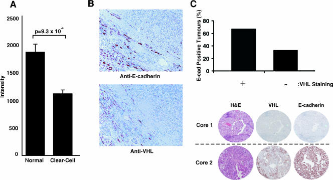
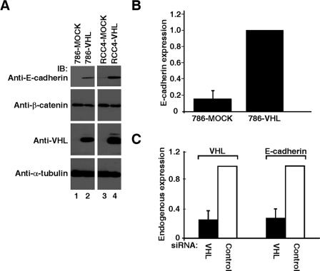
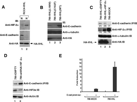
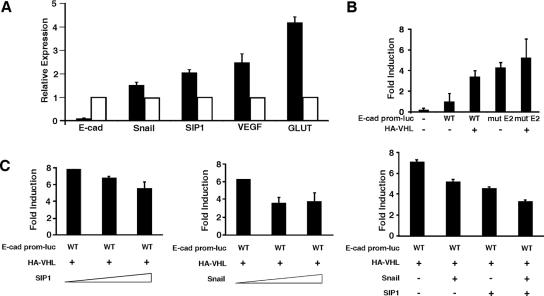
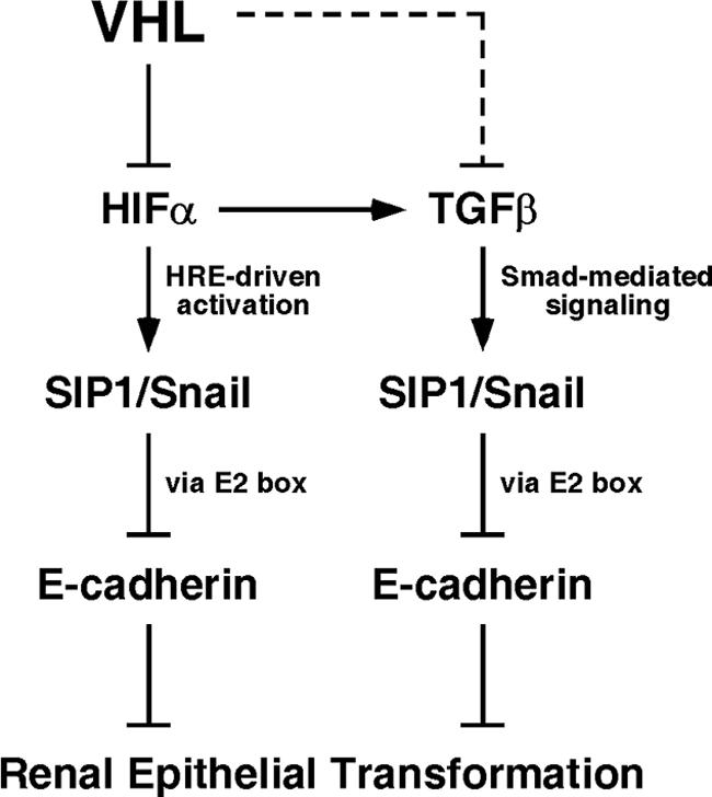
The product of the von Hippel-Lindau gene (VHL) acts as the substrate-recognition component of an E3 ubiquitin ligase complex that ubiquitylates the catalytic alpha subunit of hypoxia-inducible factor (HIF) for oxygen-dependent destruction. Although emerging evidence supports the notion that deregulated accumulation of HIF upon the loss of VHL is crucial for the development of clear-cell renal cell carcinoma (CC-RCC), the molecular events downstream of HIF governing renal oncogenesis remain unclear. Here, we show that the expression of a homophilic adhesion molecule, E-cadherin, a major constituent of epithelial cell junctions whose loss is associated with the progression of epithelial cancers, is significantly down-regulated in primary CC-RCC and CC-RCC cell lines devoid of VHL. Reintroduction of wild-type VHL in CC-RCC (VHL(-/-)) cells markedly reduced the expression of E2 box-dependent E-cadherin-specific transcriptional repressors Snail and SIP1 and concomitantly restored E-cadherin expression. RNA interference-mediated knockdown of HIFalpha in CC-RCC (VHL(-/-)) cells likewise increased E-cadherin expression, while functional hypoxia or expression of VHL mutants incapable of promoting HIFalpha degradation attenuated E-cadherin expression, correlating with the disengagement of RNA polymerase II from the endogenous E-cadherin promoter/gene. These findings reveal a critical HIF-dependent molecular pathway connecting VHL, an established "gatekeeper" of the renal epithelium, with a major epithelial tumor suppressor, E-cadherin.


Figures

FIG. 1.
FIG. 1.
Expression of E-cadherin is down-regulated in CC-RCC and correlates with VHL status. (A) 105 CC-RCC tumor samples and 12 normal kidney tissue samples were analyzed using Affymetrix HGU133 Plus 2.0 GeneChip oligonucleotide arrays. Mean E-cadherin expression and standard error were calculated, and a two-tailed Student's t test was used to determine statistical significance between the two groups. (B) Immunohistochemical staining of a representative CC-RCC with anti-E-cadherin and anti-VHL antibodies. Note the negative staining of the tumor cells (upper right in each image) with each marker, in contrast to the positive staining shown by core tubule epithelium in the adjacent nontumor renal cortex (lower left in each image) (original magnification, ×50). (C) TMAs containing 56 CC-RCC were immunostained with anti-VHL and anti-E-cad antibodies. The slides were then scanned using Aperio ScanScope and scored blind by two independent observers as being either positive or negative. Tumor cores meeting the quality standard criteria (see Materials and Methods) were considered for the correlation analysis (bar graph). Representative cores of CC-RCC on TMAs stained with anti-VHL and anti-E-cadherin antibodies are shown where the top row depicts a representative core showing negative staining for VHL and E-cadherin and the bottom row depicts a representative core showing positive staining for VHL and E-cadherin. Tumor morphology and classification were assessed using standard hematoxylin and eosin staining.
FIG. 2.
FIG. 2.
Loss of VHL results in down-regulation of E-cadherin. (A) VHL/ 786-O and RCC4 cells stably expressing wild-type VHL or empty plasmid (MOCK) were lysed, equal amounts of total cellular lysates were separated by SDS-PAGE, and immunoblotted with the indicated antibodies. Anti-α-tubulin immunoblotting was performed as an internal loading control. (B) Expression of E-cadherin was measured by quantitative real-time PCR in 786-MOCK and 786-VHL cells and normalized to U1AsnRNP1 mRNA level. The E-cadherin level in 786-VHL cells was arbitrarily set to 1.0. Error bars represent standard deviations of the relative increases in expression between the indicated cell types over three independent experiments. (C) Endogenous VHL in HEK293A cells was knocked down using VHL-specific siRNA or scrambled nontargeting control siRNA. RNA was then extracted for cDNA synthesis and endogenous transcript levels of VHL, E-cadherin, and U1AsnRNP1 measured. Error bars represent standard deviations of the relative increases between the expression of the indicated mRNA relative to its expression using control siRNA (arbitrarily set to 1.0) over three independent experiments.
FIG. 3.
FIG. 3.
Down-regulation of E-cadherin increases the migration of embryonic kidney cells and invasion of CC-RCC cells. (A) HEK293A cells were transiently transfected with a plasmid encoding the scrambled shRNA or a cocktail of four E-cadherin-specific shRNAs. Equal amounts of the whole-cell lysates were immunoprecipitated with an anti-E-cadherin antibody, resolved by SDS-PAGE, and immunoblotted with an anti-E-cadherin antibody. Equal amounts of the remaining whole-cell lysates were resolved by SDS-PAGE and immunoblotted with an anti-γ-tubulin antibody. E-cadherin signal intensities were quantified using a Kodak Image Station 2000R densitometer and normalized against the corresponding γ-tubulin signals; values are indicated in the parentheses. (B) Wounds were created 48 h posttransfection with the indicated plasmids. Percent wound closure was determined by measuring the migration of cells from the wound edge 25 h postwound scrape. Each wound measurement was taken in triplicate, and the experiment was repeated three times. (C) Line graph representing early migration profile, as indicated by percent wound closure, as measured in the experiment shown in panel B, of HEK293A cells transfected with the indicated shRNA plasmids. (D) 786-VHL cells were transiently transfected with a plasmid encoding the scrambled shRNA or a cocktail of four E-cadherin-specific shRNAs. Equal amounts of the whole-cell lysates were immunoprecipitated with an anti-E-cadherin antibody, resolved by SDS-PAGE, and immunoblotted with an anti-E-cadherin antibody. Equal amounts of the remaining whole cell lysates were resolved by SDS-PAGE and immunoblotted with an anti-hnRNP antibody. E-cadherin signal intensities were quantified using a Kodak Image Station 2000R densitometer and normalized against the corresponding hnRNP signals; results are given in parentheses. (E) 786-VHL cells were transiently transfected with the indicated plasmids as shown in panel D. Cells were counted 72 h posttransfection, and 2.5 × 10⁴ cells were seeded into BD Matrigel Invasion Chambers and incubated for 22 h. The invading cells were stained with 0.1% crystal violet, and images were captured under an inverted light microscope. Cells were counted from photographs of the membrane, and each experiment was repeated twice. The relative change in invasion was determined by counting the number of invading cells transfected with E-cadherin-specific shRNA and normalizing the value against the number of invading cells transfected with the scrambled shRNA (arbitrarily set at 1.0). Anti-E-cad, anti-E-cadherin; IP, immunoprecipitation; IB, immunoblot; Anti-Tub, anti-γ-tubulin; shE-cad, E-cadherin-specific shRNA; shScram, scrambled shRNA; T, time.
FIG. 4.
FIG. 4.
VHL regulation of E-cadherin is HIF mediated. (A) RCC4-VHL cells were maintained under normoxia (N; 21% O2) or hypoxia (H; 1% O2) for 16 h, lysed, resolved on SDS-PAGE, and immunoblotted with anti-HIF-2α, anti-E-cadherin, and anti-HA antibodies. (B) 786-O cells stably expressing HA-VHL(WT), HA-VHL(C162F), or HA-VHL(L188V) were lysed, resolved on SDS-PAGE, and immunoblotted with the indicated antibodies, where α-tubulin served as an internal loading control. (C) 786-MOCK and 786-VHL cells infected with “empty” retrovirus (786-VHL+EMPTY) or retrovirus expressing constitutively stable and functional HIF-2α(P531A) were lysed, resolved by SDS-PAGE, and immunoblotted with the indicated antibodies, where α-tubulin served as an internal loading control. Signal intensities were quantified using a Kodak Image Station 2000R densitometer and normalized against the corresponding α-tubulin signals; results are given in parentheses. Experiments were performed three times with one representative experiment presented. (D) 786-O (VHL/) subclones stably expressing pRetroSUPER-empty or pRetroSUPER-HIF2α shRNA were lysed, and comparable amounts of whole-cell extracts were immunoprecipitated and immunoblotted with an anti-E-cadherin antibody. Equal amounts of the whole-cell extracts were also resolved on SDS-PAGE and immunoblotted with anti-HIF-2α and anti-actin antibodies. Experiments were performed three times with one representative experiment presented. (E) Dual-luciferase assays were performed in 786-MOCK and 786-VHL cells transfected with the firefly luciferase construct (E-cad prom-luc) driven by the human E-cadherin promoter sequence. Cytomegalovirus-driven Renilla luciferase was used as an internal transfection control, and the firefly luciferase RLUs were normalized against Renilla luciferase RLUs. Experiments and transfections were performed in triplicate with one representative experiment presented. Error bars represent standard deviations. IP, immunoprecipitation; IB, immunoblot.
FIG. 5.
FIG. 5.
VHL-mediated transcription of E-cadherin is attenuated by Snail and SIP1 via the conserved E2 boxes. (A) Expression levels of E-cadherin, Snail, SIP1, VEGF, and GLUT-1 were measured by quantitative real-time PCR in 786-MOCK and 786-VHL cells and normalized to U1AsnRNP1 mRNA expression. Solid bars represent expression of the indicated mRNA in 786-MOCK cells relative to its expression in 786-VHL cells, which was arbitrarily set to 1.0. (B) Dual-luciferase assays were performed in U2OS cells transfected with the indicated expression plasmids. The firefly luciferase construct (E-cad prom-luc) was driven by the human E-cadherin promoter sequence (WT) or the promoter with point mutations in both E2 boxes (mutE2). Cytomegalovirus-driven Renilla luciferase was used as an internal transfection control, and the firefly luciferase RLUs were normalized against Renilla luciferase RLUs. Experiments and transfections were performed in triplicate with one representative experiment presented. Error bars represent standard deviations. (C) The experiment was performed as described in panel B with increasing concentrations of Snail and SIP1 individually mixed into HA-VHL transfection reactions at a ratio of 1:2, 2:2, and 4:2 (Snail or SIP1:HA-VHL) or equal quantities of SIP1 and Snail combined into HA-VHL transfection reactions at a ratio of 1:1 (Snail and SIP1:HA-VHL). Relative increases in induction (n-fold) were standardized to the E-cad prom-luc activity in the absence of exogenous VHL.
FIG. 6.
FIG. 6.
VHL activity is required for E-cadherin transcription. (A) ChIP using anti-RNA Pol II antibody was performed on sheared chromatin from 786-O cell lines (VHL/) that had been stably transfected with wild-type VHL (open bar) or mutant VHL(C162F) (solid bar). IP DNA was determined for the promoter and exon 10 of E-cadherin and the promoter of cyclophilin A using real-time PCR, and the value in VHL(WT) cells was arbitrarily set to 1.0. (B) RNA Pol II ChIPs were performed in 786-VHL(WT) cells exposed to 4 or 20 h of hypoxia (1% oxygen). IP DNA for exon 10 of E-cadherin was normalized to the IP DNA for the cyclophilin A promoter (left). Normoxia was arbitrarily set to 1.0. Expression of VEGF was assessed by real-time PCR as internal control for hypoxia treatment (right).
FIG. 7.
FIG. 7.
VHL gatekeeper's pathway in renal epithelium. See text for details.

Similar articles

Cited by

References

    1. Affymetrix. 2003. GeneChip expression analysis technical manual. Affymetrix, Santa Clara, CA.
    1. Ananth, S., B. Knebelmann, W. Gruning, M. Dhanabal, G. Walz, I. Stillman, and V. Sukhatme. 1999. Transforming growth factor beta1 is a target for the von Hippel-Lindau tumor suppressor and a critical growth factor for clear cell renal carcinoma. Cancer Res. 59:2210-2216. - PubMed
    1. Baba, M., S. Hirai, S. Kawakami, T. Kishida, N. Sakai, S. Kaneko, M. Yao, T. Shuin, Y. Kubota, M. Hosaka, and S. Ohno. 2001. Tumor suppressor protein VHL is induced at high cell density and mediates contact inhibition of cell growth. Oncogene 20:2727-2736. - PubMed
    1. Baba, M., S. Hirai, H. Yamada-Okabe, K. Hamada, H. Tabuchi, K. Kobayashi, K. Kondo, M. Yoshida, A. Yamashita, T. Kishida, N. Nakaigawa, Y. Nagashima, Y. Kubota, M. Yao, and S. Ohno. 2003. Loss of von Hippel-Lindau protein causes cell density dependent deregulation of CyclinD1 expression through hypoxia-inducible factor. Oncogene 22:2728-2738. - PubMed
    1. Batlle, E., E. Sancho, C. Franci, D. Dominguez, M. Monfar, J. Baulida, and A. Garcia De Herreros. 2000. The transcription factor snail is a repressor of E-cadherin gene expression in epithelial tumour cells. Nat. Cell Biol. 2:84-89. - PubMed

Publication types

MeSH terms